Can a Resting EEG Tell Us How Well the Brain Is Ready to Think?
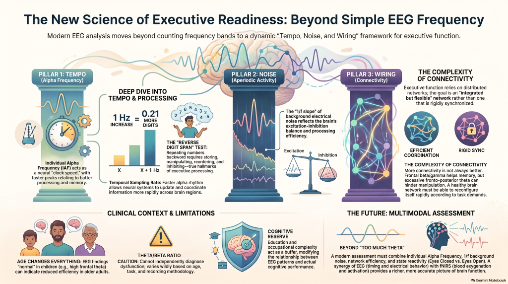 What alpha frequency, brain “noise,” connectivity and qEEG may tell us about attention and executive function
Why can one person remain focused, manipulate several pieces of information in their mind and quickly adapt to changing demands, while another struggles even when both are equally motivated?
Part of the answer may lie in the brain’s baseline state before a task even begins.
A major 2026 systematic review published in the Journal of Clinical Medicine examined 63 studies investigating whether resting-state EEG could predict differences in executive function. The studies covered working memory, attention, inhibition, decision-making, processing speed and cognitive flexibility across different age groups.
The emerging message is important:
There is probably no single “attention wave” or EEG number that tells us how well the brain works.
Instead, executive performance appears to depend on a combination of three broad characteristics:
Tempo. Noise. Wiring.
What are executive functions?
Executive functions are the brain’s control systems.
They allow us to:
- keep a goal in mind,
- resist distraction,
- hold information temporarily,
- manipulate information mentally,
- stop inappropriate responses,
- switch between tasks,
- make decisions,
- monitor mistakes,
- and sustain attention over time.
Working memory, inhibitory control and cognitive flexibility are partly distinct processes, but they also share a common ability to maintain goals and resist interference.
These functions become particularly important in conditions where patients complain of:
- poor concentration,
- forgetfulness,
- procrastination,
- distractibility,
- impulsivity,
- inconsistent productivity,
- mental fatigue,
- or difficulty organising complex tasks.
But measuring executive function objectively is difficult. Even commonly used cognitive tests contain several different cognitive demands at the same time.
EEG offers another window into the problem.
The resting brain is not really “doing nothing”
During a resting-state EEG, a person is awake but not performing a specific cognitive task.
Traditionally, EEG analysis has focused heavily on the quantity of activity within familiar frequency bands:
Delta → Theta → Alpha → Beta → Gamma
But the new review suggests that this is an incomplete way of looking at the brain.
The most informative EEG profile may instead involve:
1. Tempo
How rapidly the brain’s dominant rhythms operate.
2. Noise
The organisation of the broadband background electrical signal.
3. Wiring
How efficiently different brain regions communicate with each other.
Together, these may provide a physiological picture of what the authors describe as “executive readiness”—the brain’s baseline capacity to deploy cognitive control when required.
1. TEMPO: How fast is the brain’s alpha rhythm?
Alpha activity usually falls around 8–13 Hz.
Most people are familiar with measuring the amount of alpha activity.
However, the review suggests something more interesting:
The speed of the alpha rhythm may sometimes matter more than the amount of alpha.
This is measured using the Individual Alpha Frequency (IAF) or Peak Alpha Frequency (PAF).
Consider two individuals.
One person’s strongest alpha activity may occur around:
8.5 Hz
while another person’s peak may occur around:
10.5 Hz.
Both may technically have “normal alpha.”
But their underlying temporal organisation may be different.
Across several studies, a faster alpha peak was associated with better processing speed, interference control, sustained attention and demanding forms of working memory.
One particularly interesting study involving 550 participants found that every 1 Hz increase in frontal alpha peak frequency predicted approximately 0.21 additional digits on reverse digit span.
Why is reverse digit span important?
Repeating:
7 – 2 – 9
as:
7 – 2 – 9
mainly requires storage.
But repeating it backwards:
9 – 2 – 7
requires the brain to:
store → manipulate → reorder → inhibit the original sequence → respond.
That is much closer to true executive processing.
Interestingly, resting EEG associations often become clearer when tasks are sufficiently demanding. Simple low-load tasks frequently produce weak or absent relationships.
Alpha frequency may represent something like neural “clock speed”
This does not mean that a higher alpha frequency is always better.
Age, development and cognitive reserve all change its interpretation.
But one useful model is that alpha frequency represents something similar to the brain’s temporal sampling rate.
A faster rhythm could potentially allow neural systems to update and coordinate information more rapidly.
This may partly explain why alpha frequency appears more consistently related to some executive abilities than simple alpha power.
2. NOISE: The hidden background underneath EEG rhythms
A conventional EEG spectrum contains familiar peaks.
But beneath those peaks is a broadband background signal that decreases with increasing frequency.
This is called the aperiodic component, often described using a 1/f slope or exponent and an offset.
Modern EEG analysis increasingly separates:
Periodic activity
from
Aperiodic activity
because failing to separate them can lead to misleading conclusions.
For example, what appears to be “increased theta” could sometimes partly reflect a change in the overall spectral slope rather than a true increase in a specific theta oscillation.
The review identifies this as an important limitation of conventional band-power interpretation.
What does the 1/f slope mean?
The biological interpretation remains an active area of research.
However, aperiodic EEG characteristics have been associated with factors including:
- neural excitation–inhibition balance,
- population firing behaviour,
- arousal,
- processing efficiency,
- and age-related neural change.
Across studies reviewed in the paper, steeper aperiodic slopes and appropriate offsets were often associated with faster processing and stronger executive performance, although the relationship was not universal and could change with age and cognitive reserve.
This means that simply reporting:
“Theta is high.”
or
“Alpha is low.”
may sometimes be physiologically incomplete.
Modern qEEG interpretation increasingly needs to ask:
Is this a genuine oscillatory change, or has the entire background spectrum changed?
3. WIRING: How efficiently are brain regions communicating?
Perhaps the most important conclusion of the review is that network organisation may sometimes predict executive performance better than local EEG power.
The brain does not perform executive functions using one electrode or one cortical region.
Attention and working memory require communication across distributed networks.
Therefore, measures of:
- coherence,
- phase relationships,
- connectivity,
- global efficiency,
- local efficiency,
- characteristic path length,
- clustering,
- and network flexibility
may provide information that simple power measurements cannot.
The review repeatedly found that efficient network organisation was associated with faster and more reliable executive performance.
More connectivity is not necessarily better
This is an important point.
It is tempting to assume:
low connectivity = bad
and:
high connectivity = good.
The evidence does not support such a simple interpretation.
For working memory, stronger within-frontal beta and gamma coherence was associated with better maintenance and sequencing.
But excessive fronto-posterior theta coherence was associated with poorer working-memory manipulation and updating.
In other words:
The brain needs coordination without becoming rigidly synchronized.
A network that is strongly connected but unable to reconfigure may actually perform worse.
The ideal brain network may be integrated but flexible
Graph-theoretical studies provide another way of analysing this.
Better-performing networks often demonstrate:
- shorter communication paths,
- greater efficiency,
- useful local specialization,
- and the ability to reorganise according to demand.
But excessive synchronization can become counterproductive.
For example, rigid high-beta networks were associated with poorer performance and reduced task-related network reconfiguration, while highly synchronized alpha/gamma networks could be behaviourally slower.
Therefore:
Healthy brain function may depend less on maximal connectivity and more on efficient, flexible connectivity.
Working memory: one of the clearest EEG relationships
Working memory is our ability to temporarily hold and manipulate information.
The review suggests that better manipulation-heavy working memory tends to be associated with:
- faster frontal alpha frequency,
- appropriate frontal beta/gamma coherence,
- efficient network integration,
- and less excessive long-range slow-frequency synchronization.
Meanwhile, excessive fronto-posterior theta connectivity may interfere particularly with sequencing and manipulation.
This may help explain why someone can appear to have adequate simple memory yet struggle when asked to:
- mentally calculate,
- reorganise information,
- follow multiple instructions,
- plan several steps ahead,
- or remember something while simultaneously doing another task.
Inhibitory control: stopping yourself is a separate brain function
Executive control is not a single ability.
Suppressing a motor response is different from:
- ignoring distraction,
- resisting emotional interference,
- maintaining attention,
- or suppressing an automatic thought.
Interestingly, higher resting beta activity is not always advantageous.
In motor and inhibitory networks, greater beta power and stronger beta connectivity have sometimes been associated with longer stop-signal reaction times, suggesting poorer response inhibition.
Again:
“More beta” does not automatically mean better attention.
What about the famous theta/beta ratio?
The theta/beta ratio, or TBR, has historically received considerable attention, particularly in discussions around attention and ADHD.
The new review urges considerable caution.
Higher frontal theta/beta ratios were associated in some studies with:
- poorer reversal learning,
- riskier decision-making,
- less advantageous choices,
- altered emotional inhibition,
- and greater deterioration of attention under stress.
Lower ratios were sometimes associated with better attentional orienting.
But these relationships varied substantially depending on:
- age,
- task,
- cognitive reserve,
- recording methodology,
- and population.
The authors therefore conclude that simple ratios such as theta/beta cannot independently diagnose executive dysfunction.
This is particularly important when interpreting qEEG in ADHD.
A theta/beta ratio may contribute information.
It should not become the diagnosis.
Age changes everything
One of the strongest messages in the review is that the same EEG finding can have a different meaning at different ages.
For example:
In children
Higher frontal theta can sometimes accompany better working memory.
In older adults
Greater slow-wave activity and slowing of alpha are more often associated with reduced cognitive efficiency.
The relationship between theta/alpha ratios and cognition can even reverse across age groups.
Therefore, interpreting a child’s EEG using an adult framework—or an older adult’s EEG using a young-adult framework—is scientifically inappropriate.
Cognitive reserve also matters
Two people with similar EEG patterns may perform differently because the brain is shaped by years of experience.
Factors associated with cognitive reserve include:
- education,
- occupational complexity,
- intellectually stimulating activities,
- social engagement,
- and broader lifetime experience.
The review found that cognitive reserve could modify relationships between EEG and executive function.
Higher reserve was associated with more flexible state-dependent connectivity changes, including stronger differences between eyes-open and eyes-closed states.
This means that EEG values should not be interpreted independently of the individual.
Eyes-open and eyes-closed EEG provide different information
Resting EEG is commonly recorded with the eyes closed.
But the transition between eyes closed and eyes open is itself informative.
Opening the eyes normally suppresses posterior alpha activity and reorganises network activity.
Therefore, comparing:
Eyes closed → Eyes open
can provide information about how dynamically the brain responds to changing environmental demands.
The paper argues for recording both states rather than treating them as interchangeable.
Beyond frequency bands: microstates and temporal dynamics
Modern EEG can also analyse the brain at much shorter timescales.
EEG microstates
The scalp electrical pattern briefly settles into relatively stable configurations before switching to another.
Their:
- duration,
- occurrence,
- coverage,
- and transition pattern
may reveal aspects of large-scale brain-state organisation.
However, the review finds that microstate measures vary substantially with age and cannot yet be treated as simple markers of specific executive abilities.
How “sticky” is brain activity?
Researchers can also examine long-range temporal correlations.
Instead of asking:
“How much theta does this brain produce?”
we can ask:
“How strongly does the current activity depend on what the brain was doing moments earlier?”
Very persistent activity may reflect a network that is less flexible.
In one study, stronger long-range temporal correlations in theta and delta activity were associated with poorer working-memory ability even after accounting for EEG power.
This reinforces a recurring idea throughout the review:
Good cognition may require stability without rigidity.
Speed and accuracy must be separated
Another important clinical lesson is that “better performance” is not one thing.
Some brain patterns may support:
faster responses
while others favour:
more stable and accurate responses.
For example, certain posterior alpha patterns may support stable monitoring but result in slower reaction times.
Therefore, cognitive testing should ideally examine:
- average reaction time,
- response variability,
- omission errors,
- commission errors,
- accuracy,
- response inhibition,
- and possibly evidence-accumulation measures.
The review specifically recommends separating speed from accuracy rather than collapsing both into one cognitive score.
What does this mean for qEEG assessment?
The paper suggests that a modern qEEG assessment should increasingly move beyond simply asking:
Is theta high?
Is alpha low?
Is beta excessive?
A more sophisticated approach asks:
Tempo
What is the person’s individual alpha frequency?
Noise
What does the aperiodic 1/f background look like?
Wiring
How efficiently are relevant brain regions communicating?
Flexibility
Can the network reconfigure appropriately?
Reactivity
How does the brain change from eyes closed to eyes open?
Behaviour
Do these physiological findings correspond to objectively measurable cognitive performance?
This creates a much more meaningful model:
EEG physiology + cognitive performance + clinical history
rather than attempting to diagnose a person from an isolated EEG number.
Can EEG diagnose ADHD or executive dysfunction?
Not by itself.
This is one of the most important conclusions of the review.
Resting EEG may provide information about an individual’s baseline executive readiness, but the authors explicitly caution against using a single frequency band or ratio as a stand-alone diagnostic biomarker.
The evidence currently supports EEG more strongly for:
characterisation, stratification, research and potentially longitudinal monitoring
than for replacing a clinical diagnosis.
This distinction matters.
A biological correlate is not automatically a diagnostic test.
And a diagnostic correlate is not automatically a treatment target.
What would a high-quality resting EEG require?
The review recommends that biomarker-quality investigations include at least approximately:
- 5–6 minutes of eyes-closed EEG,
- an eyes-open recording,
- 32 or more channels,
- appropriate artifact removal,
- careful monitoring for drowsiness,
- separation of periodic and aperiodic activity,
- and analysis beyond simple band power.
For future research, the authors propose even more rigorous high-density and multimodal protocols.
EEG and fNIRS may be particularly complementary
One especially interesting future direction is combining EEG with functional near-infrared spectroscopy — fNIRS.
EEG provides extraordinarily fast information about electrical brain activity.
fNIRS measures changes in cortical oxygenated and deoxygenated haemoglobin associated with regional brain activation.
The review identifies simultaneous EEG–fNIRS as a practical portable approach for studying executive networks and recommends exploring whether the two modalities together can provide better prediction and monitoring than either alone.
Conceptually:
EEG tells us about timing and electrical network behaviour.
fNIRS tells us about cortical haemodynamic activation.
Combining electrophysiological and haemodynamic information could therefore provide a richer picture of brain function.
But this remains an emerging research direction rather than a validated diagnostic shortcut.
The future of EEG is probably not a single biomarker
Perhaps the strongest lesson from this review is that the search for one perfect EEG biomarker may itself be misguided.
The brain appears to work more like a dynamic network.
A stronger executive profile may involve:
faster appropriate alpha timing
a favourable aperiodic background
efficient network communication
limited pathological over-synchronization
appropriate brain-state flexibility
measurable cognitive performance.
The review therefore shifts the question from:
“Which EEG frequency is abnormal?”
to:
“How is this person’s brain organised to deploy attention and executive control?”
That is a much more useful question.
And it may represent the direction in which objective cognitive neuroscience is moving.
Bottom line
Resting-state EEG can tell us something meaningful about the brain’s readiness for executive control, but it cannot tell the whole story.
The strongest evidence does not support interpreting one electrode, one frequency or one ratio in isolation.
Instead, executive function appears to emerge from the interaction between:
Tempo + Noise + Wiring + Flexibility + Behaviour
The future of qEEG is therefore likely to be multimodal and multimetric—combining sophisticated EEG analysis with objective cognitive testing, clinical assessment and, increasingly, complementary technologies such as fNIRS.
That approach is scientifically more cautious.
It is also far more interesting than simply looking for “too much theta.”