Brain Mapping for ADHD: What qEEG Can Tell Us About the ADHD Brain
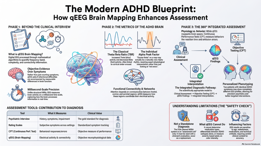 Can we actually see ADHD in the electrical activity of the brain?
Attention-Deficit/Hyperactivity Disorder (ADHD) is diagnosed clinically. There is no single blood test, MRI scan or EEG pattern that can independently prove that a person has ADHD. However, advances in quantitative electroencephalography (qEEG), commonly called brain mapping, are making it possible to examine the neurophysiology underlying attention, arousal and information processing in considerably greater detail.
Rather than asking only, “Does this person have enough symptoms for ADHD?”, modern assessment can ask an additional question:
“Is there objective evidence that this person’s attentional difficulties are accompanied by measurable differences in brain function?”
That is where qEEG becomes interesting.
What Is Brain Mapping or qEEG?
A conventional EEG records tiny electrical signals generated by neuronal activity using electrodes placed on the scalp.
Quantitative EEG takes this a step further.
The digital EEG signal is processed mathematically to quantify characteristics such as:
- delta, theta, alpha and beta activity
- absolute and relative power
- dominant frequencies
- theta/beta relationships
- asymmetry between brain regions
- coherence and functional connectivity
- network characteristics
These measurements can then be displayed as topographic brain maps, allowing patterns of electrical activity across different scalp regions to be visualised.
The review by Popa and colleagues describes qEEG as digital EEG processed using mathematical algorithms to extract frequency-band, complexity, connectivity and network information. Importantly, the authors emphasise that qEEG should provide additional objective information alongside other diagnostic evaluations rather than function as an immediate stand-alone diagnosis.
Why Is Brain Mapping Particularly Interesting in ADHD?
ADHD is a neurodevelopmental disorder involving much more than simply “poor concentration.”
The clinical phenotype can involve abnormalities in:
- sustained attention
- response inhibition
- vigilance
- working memory
- executive control
- reward processing
- arousal regulation
- reaction-time consistency
- impulse control
These functions depend on distributed brain networks rather than one isolated “attention centre.”
This is one reason electrophysiology is attractive in ADHD: EEG measures brain activity on a millisecond time scale and therefore provides a different kind of information from structural imaging such as MRI.
But there is an important distinction.
qEEG does not produce a photograph of ADHD. It measures electrical characteristics that may be associated with the neurophysiology of ADHD.
The Classical ADHD Brain-Wave Pattern
For decades, one of the best-known EEG findings in ADHD has been an alteration in the balance between slower and faster brain-wave activity.
The frequently discussed pattern is:
↑ Theta activity
↓ Beta activity
→ Increased Theta/Beta Ratio
Theta activity is generally concentrated in approximately the 4–8 Hz range, whereas beta represents faster electrical activity associated with alert cognitive processing.
Early ADHD studies therefore proposed that some individuals with ADHD demonstrate a state of relative cortical under-arousal or delayed cortical maturation, producing excessive slow-wave activity relative to faster activity.
The review supplied for this article notes that children and adults with ADHD have been reported to show increased theta and delta power, while reduced beta power has been reported particularly in adolescents. It also describes historical work showing an elevated theta/beta ratio at Cz in ADHD, while importantly warning that this pattern is not specific to ADHD and cannot simply be generalised to every patient.
This last point has become increasingly important.
The Theta/Beta Ratio Is Useful—but ADHD Is More Complicated Than One Number
The theta/beta ratio (TBR) became one of the most widely investigated electrophysiological markers in ADHD.
There is enough biological signal in this measurement that the US FDA classified the NEBA system, which measured theta/beta ratio at the Cz electrode in children and adolescents aged 6–17 years, as a Class II neuropsychiatric EEG assessment aid.
However, the FDA indication is very specific: NEBA was designed to be used together with a clinician’s evaluation, either as confirmatory support or to indicate that further investigation may be useful. The FDA documentation explicitly states that it must not be used as a stand-alone ADHD diagnostic test.
That distinction remains crucial.
A high theta/beta ratio does not mean:
“This patient definitely has ADHD.”
And a normal theta/beta ratio does not mean:
“This patient definitely does not have ADHD.”
ADHD is neurobiologically heterogeneous.
There May Be Several Electrophysiological Types of ADHD
One of the most important developments in modern ADHD electrophysiology is the recognition that there may not be one universal “ADHD EEG.”
Recent research has challenged the idea that every person with ADHD should have an elevated theta/beta ratio.
For example, Kiiski and colleagues examined resting EEG in adults with ADHD and found that patterns involving delta, theta, low-alpha and beta spectral power differentiated groups better than theta/beta ratio alone; TBR itself did not successfully classify adult ADHD.
More recent work has gone further.
Boxum and colleagues analysed 417 children and adolescents with ADHD aged 6–18 years and demonstrated that an apparently high theta/beta ratio can arise through different electrophysiological mechanisms. One subgroup showed genuinely increased TBR, whereas another showed a slower alpha peak frequency spilling into the conventional theta range. The EEG subgroups were not reliably associated with baseline ADHD symptom severity.
This is an important conceptual shift.
ADHD brain mapping is moving from:
“Does this person have an elevated theta/beta ratio?”
towards:
“What electrophysiological phenotype does this individual show?”
That is much closer to the idea of precision psychiatry.
Looking Beyond Theta/Beta Ratio
Modern qEEG analysis can examine considerably more than a single ratio.
1. Absolute and Relative Spectral Power
Instead of merely dividing theta by beta, qEEG can examine the amount and distribution of:
Delta → Theta → Alpha → Beta
across multiple brain regions.
The spatial distribution matters.
For example, excessive frontal theta activity may carry different neurophysiological implications from diffuse slow-wave activity.
A 2025 study using multidimensional EEG analysis in children with ADHD found differences involving theta, alpha and beta power together with alterations in functional connectivity. The authors’ machine-learning framework extracted power, complexity and connectivity information rather than relying on one EEG marker.
2. Individual Alpha Peak Frequency
This is becoming increasingly important.
If a person’s alpha rhythm is naturally slower, part of that alpha activity can be counted mathematically as theta.
The brain map may consequently appear to demonstrate “excess theta” when the actual phenomenon is slow alpha.
This is one reason brain mapping should be interpreted physiologically rather than simply by looking for red areas on a colour map.
3. Functional Connectivity
The brain does not operate as isolated electrodes.
Attention depends on communication between networks involving frontal, central, parietal and subcortical systems.
qEEG can therefore examine measures of functional connectivity—how electrical activity recorded from different regions varies together.
Contemporary ADHD research increasingly investigates these network-level abnormalities rather than searching for a single abnormal frequency. Recent multidimensional EEG research has demonstrated differences in functional connectivity across theta, alpha, beta and other frequency ranges in children with ADHD.
4. Asymmetry
Electrical activity can also be compared between:
- left and right hemispheres
- frontal regions
- central regions
- parietal regions
- anterior and posterior networks
These measures may provide additional information about functional organisation, although no single asymmetry pattern is diagnostic of ADHD.
5. Brain Activity During Cognitive Tasks
Resting EEG asks:
“What is the brain doing when the person is sitting quietly?”
Task-related EEG asks a potentially more relevant ADHD question:
“What happens when the brain actually has to sustain attention, inhibit responses or process information?”
A 2024 study examining children with ADHD found relationships between theta/beta ratio and measures obtained during the Conners Continuous Performance Test, illustrating why combining electrophysiology with objective attention testing may be more informative than interpreting either measure in isolation.
qEEG and Continuous Performance Testing Measure Different Things
This distinction is extremely useful clinically.
A Continuous Performance Test (CPT) primarily measures behaviour during an attention-demanding task:
- omission errors
- commission errors
- reaction time
- variability in reaction time
- sustained attention
- inhibitory control
qEEG measures electrophysiology.
Rating scales measure symptoms and functioning.
The psychiatric interview evaluates:
- developmental history
- age of onset
- impairment
- symptoms across settings
- differential diagnosis
- comorbidity
These are complementary pieces of information.
A modern ADHD assessment can therefore be conceptualised as:
Clinical phenotype + behavioural phenotype + neurophysiological phenotype
rather than expecting one test to answer everything.
Why Clinical Assessment Still Comes First
Major ADHD guidelines continue to define ADHD clinically.
The American Academy of Pediatrics recommends establishing ADHD symptoms and functional impairment in more than one setting, obtaining information from relevant observers and excluding alternative explanations.
That is essential because poor attention can occur in many other conditions, including:
- anxiety disorders
- depression
- autism spectrum disorder
- specific learning disorders
- sleep deprivation
- obstructive sleep apnoea
- substance use
- medication effects
- intellectual or developmental disorders
- neurological disorders
Even the FDA-cleared EEG-based NEBA approach required a clinical ADHD evaluation before EEG information was incorporated into the diagnostic pathway.
So the scientifically appropriate model is not:
qEEG → diagnosis
but rather:
Clinical assessment → objective testing → qEEG findings → integrated interpretation
What Can qEEG Add to an ADHD Assessment?
Used carefully, brain mapping can provide several potentially valuable layers of information.
Objective neurophysiological data
It transforms electrical brain activity into measurable variables rather than relying exclusively on symptom descriptions.
Identification of electrophysiological heterogeneity
Two patients meeting the same clinical criteria for ADHD may have very different EEG profiles.
Examination of arousal regulation
Patterns involving theta, alpha and beta activity can provide clues about cortical activation and vigilance.
Integration with cognitive testing
Comparing qEEG findings with CPT performance may help determine whether neurophysiological abnormalities correspond with objectively demonstrated problems in attention or response control.
Baseline for longitudinal assessment
Repeated measurements performed under sufficiently standardised conditions can potentially examine neurophysiological changes over time.
Neurofeedback planning and research
Electrophysiological phenotyping may become particularly relevant when neurofeedback protocols are being considered. Recent work, for example, has suggested that TBR may be more useful for stratifying particular neurofeedback approaches than for diagnosing ADHD itself.
What qEEG Cannot Do
This is equally important.
A colourful brain map should never be interpreted as diagnostic proof.
qEEG currently cannot reliably determine on its own:
- whether someone definitely has ADHD
- whether ADHD is predominantly inattentive or hyperactive
- which medication will definitely work
- whether a child requires stimulant medication
- the severity of ADHD solely from colour intensity
- whether every abnormality detected is caused by ADHD
EEG is influenced by factors including age, wakefulness, medications, recording conditions, electrode characteristics and artifacts. The Popa review specifically highlights biological, technical and methodological variability and concludes that qEEG is best treated as complementary objective information rather than an immediate diagnostic instrument.
Brain Mapping Is Not an MRI
Patients often confuse the two.
MRI primarily provides anatomical and structural information.
EEG/qEEG measures electrical brain activity.
Brain mapping therefore tells us much more about when and how electrical activity is occurring than about the anatomical structure of the brain.
It is also non-invasive. The electrodes placed on the scalp record electrical signals; they do not send electricity into the brain.
The FDA’s review of the NEBA EEG assessment system similarly characterised EEG acquisition as a non-invasive procedure.
Why Brain Mapping May Become More Important in the Future
The next generation of ADHD biomarkers will probably not consist of one electrode, one frequency band or one ratio.
Research is increasingly moving toward combinations of:
spectral power + connectivity + signal complexity + cognitive performance + machine learning + clinical phenotype
A 2025 study using machine-learning analysis of paediatric EEG, for example, combined 206 power, entropy and connectivity features to distinguish its research ADHD and control datasets. Such results are promising, but highly accurate algorithms derived from research datasets still require independent clinical validation before they can be treated as diagnostic tests in everyday psychiatry.
The direction of travel is nevertheless important.
Psychiatry is gradually moving from:
symptoms alone
towards:
symptoms + cognition + physiology + longitudinal measurement.
ADHD is likely to be one of the disorders in which this transition occurs earliest.
A 360-Degree Approach to ADHD Assessment
The most useful role for brain mapping is therefore not to replace psychiatric assessment but to strengthen it.
At the Attention Clinic in Chennai, my approach to ADHD assessment is built around integrating multiple sources of information rather than relying on a single questionnaire or test.
Depending on age and clinical requirements, assessment can incorporate:
**Detailed psychiatric and developmental assessment
- Standardised ADHD rating scales
- Objective Continuous Performance Testing
- qEEG / brain mapping
- Evaluation of comorbidities and alternative explanations
- Integrated clinical interpretation**
When required, psychological interventions and ADHD-focused therapy can also form part of comprehensive management.
The aim is straightforward:
Not merely to label ADHD—but to understand the individual patient’s attentional phenotype as objectively and comprehensively as current evidence allows.
Frequently Asked Questions
Can a brain map diagnose ADHD?
Not by itself. qEEG can provide objective electrophysiological information that may support clinical assessment, but ADHD remains a clinical diagnosis. Even the FDA-cleared NEBA EEG assessment aid was specifically authorised only as an adjunct to clinician evaluation.
Does everyone with ADHD have a high theta/beta ratio?
No. Although elevated TBR has historically been associated with ADHD, modern studies show substantial neurophysiological heterogeneity. Some individuals have other spectral abnormalities, some have slow alpha activity, and others may not demonstrate the classical pattern at all.
Is brain mapping painful?
No. EEG is non-invasive. Electrodes placed on the scalp record naturally occurring electrical activity.
Is qEEG useful in adult ADHD?
Potentially, but adult patterns may differ from those found in children. Research in adults has found abnormalities in spectral power while failing to support theta/beta ratio alone as a sufficiently reliable diagnostic biomarker.
Is qEEG the same as a Continuous Performance Test?
No. A CPT objectively measures attention and response behaviour. qEEG measures electrical brain activity. Combining behavioural, clinical and neurophysiological information can provide a much richer assessment than treating any one test as definitive.
Can qEEG select the correct ADHD medication?
At present, qEEG should not be marketed as a test that reliably tells us which ADHD medicine a particular patient should receive. Pharmacological decisions still require clinical assessment, treatment history, comorbidity, safety considerations and monitoring.
ADHD Brain Mapping in Chennai
For children, adolescents and adults in whom ADHD is suspected—particularly when the diagnosis is complex or when a more objective assessment is desired—a structured evaluation can go beyond questionnaires alone.
Dr. Srinivas Rajkumar T
Senior Consultant Psychiatrist
Attention Clinic – Comprehensive 360° ADHD Care
Apollo Clinic, Velachery, Chennai
Opposite Phoenix Marketcity
The clinic focuses on structured ADHD evaluation integrating clinical assessment with objective attention testing and neurophysiological assessment where appropriate.
The future of ADHD assessment is unlikely to be clinical judgement versus technology. It will be clinical judgement strengthened by better technology.
Selected References
- Popa LL, Dragos H, Pantelemon C, Rosu OV, Strilciuc S. The Role of Quantitative EEG in the Diagnosis of Neuropsychiatric Disorders. J Med Life. 2020;13(1):8–15. doi:10.25122/jml-2019-0085.
- US Food and Drug Administration. De Novo Classification Request for Neuropsychiatric EEG-Based Assessment Aid for ADHD (NEBA) System. Product code NCG; Class II.
- Kiiski H, Bennett M, Rueda-Delgado LM, et al. EEG spectral power, but not theta/beta ratio, is a neuromarker for adult ADHD. Eur J Neurosci. 2020;51:2095–2109. doi:10.1111/ejn.14645.
- Wang TS, Wang SS, Wang CL, Wong SB. Theta/beta ratio in EEG correlated with attentional capacity assessed by Conners Continuous Performance Test in children with ADHD. Front Psychiatry. 2024;14:1305397. doi:10.3389/fpsyt.2023.1305397.
- Boxum M, Voetterl H, van Dijk H, et al. Challenging the Diagnostic Value of Theta/Beta Ratio: Insights From an EEG Subtyping Meta-Analytical Approach in ADHD. Appl Psychophysiol Biofeedback. 2025;50:655–666. doi:10.1007/s10484-024-09649-y.
- Mao Y, Qi X, He L, et al. Advanced machine learning techniques reveal multidimensional EEG abnormalities in children with ADHD: a framework for automatic diagnosis. Front Psychiatry. 2025;16:1475936. doi:10.3389/fpsyt.2025.1475936.