Brain Mapping in Schizophrenia and Psychosis: What qEEG, Gamma Synchrony, MMN and P300 Reveal
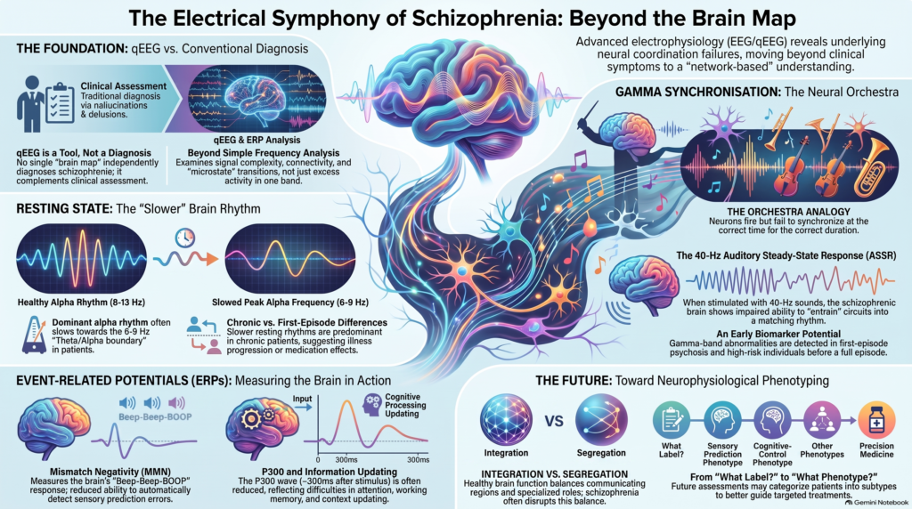 Can psychosis be seen in the electrical activity of the brain?
Schizophrenia is usually recognised clinically through disturbances involving perception, thought, cognition, motivation and behaviour. Yet beneath hallucinations, delusions, thought disorder and negative symptoms lies another level of dysfunction: the timing and coordination of neuronal networks.
This makes schizophrenia one of the most fascinating disorders for electroencephalography (EEG), quantitative EEG (qEEG) and event-related potential research.
Electrophysiological studies have identified abnormalities involving:
- increased slow-frequency activity
- altered theta/alpha organisation
- slowing of peak alpha frequency
- abnormal gamma synchronisation
- disrupted functional connectivity
- reduced mismatch negativity (MMN)
- abnormal P300 responses
- altered error and inhibitory-control processing
- abnormal EEG microstates
But the first principle is essential:
There is currently no qEEG brain map that independently diagnoses schizophrenia.
The 2020 qEEG review used throughout this brain-mapping series was appropriately cautious: although qEEG had been investigated in schizophrenia, its psychiatric application was classified at that time among experimental uses without established evidence for routine clinical diagnosis.
That does not mean the electrophysiology is unimportant.
In fact, schizophrenia may be one of the disorders in which EEG tells us the most interesting things about how neural networks fail to coordinate information.
What Does qEEG Measure?
Conventional EEG records tiny fluctuations in electrical potential produced predominantly by synchronised postsynaptic activity in cortical neuronal populations.
Quantitative EEG applies mathematical analysis to this signal.
It can examine:
Spectral power
How much electrical activity occurs within delta, theta, alpha, beta and gamma ranges?
Peak frequency
At what frequency is the dominant rhythm actually oscillating?
Asymmetry
Are corresponding brain regions functioning differently?
Coherence and connectivity
How consistently are different regions synchronising?
Phase relationships
Is the timing of communication between regions preserved?
Source localisation
Which cortical regions may be contributing to an observed signal?
Microstates
How does the brain transition between brief, relatively stable configurations of large-scale electrical activity?
The uploaded review emphasises that modern qEEG goes beyond simple frequency analysis to include signal complexity, connectivity and network analysis.
This is especially relevant to schizophrenia because schizophrenia increasingly appears to involve disordered coordination between neural systems rather than a defect in one isolated brain region. Electrophysiological studies have demonstrated abnormalities in oscillatory synchronisation and phase coherence across multiple paradigms.
The Classical Resting qEEG Finding: A Slower Brain Rhythm
One of the oldest EEG observations in schizophrenia is an alteration in the balance between slower and faster frequencies.
At the group level, resting EEG studies have often reported:
↑ Delta / theta activity
and/or
↓ or slower alpha activity
But modern analysis shows that the phenomenon is more complicated than simply saying:
“Schizophrenia causes excess theta.”
A 2024 study by Nakhnikian and colleagues used data-driven spectral decomposition rather than imposing conventional frequency bands in advance. In 39 patients with chronic schizophrenia and 36 matched controls, peak alpha frequency was significantly reduced, and the investigators identified a schizophrenia-associated spectral component in the 6–9 Hz theta/alpha region, with source distribution particularly involving prefrontal and parahippocampal areas.
That finding is particularly interesting.
1. The Theta/Alpha Boundary May Matter More Than “High Theta”
Traditional qEEG divides activity into predefined bins:
Theta: approximately 4–8 Hz
Alpha: approximately 8–13 Hz
But biology does not necessarily respect those arbitrary boundaries.
If a person’s dominant alpha rhythm becomes slower, activity that previously occurred around 9–10 Hz may shift toward 7–9 Hz.
A conventional brain map might therefore report:
“increased theta”
when part of the phenomenon may actually reflect:
a slowing of the dominant alpha-generating network.
The 2024 spectral-decomposition study is important precisely because it identified a specific 6–9 Hz theta/alpha component rather than treating all theta activity as one biological phenomenon.
This illustrates why sophisticated qEEG interpretation should move beyond simply asking:
“Which colour is abnormal?”
and instead ask:
“What physiological process produced this frequency distribution?”
2. Peak Alpha Frequency in Schizophrenia
Alpha rhythm is usually prominent during relaxed wakefulness and participates in:
- attentional regulation
- sensory gating
- cortical inhibition
- information routing
- coordination of large-scale networks
A reduction in peak alpha frequency has repeatedly attracted attention in schizophrenia research. In the 2024 study described above, peak alpha frequency was significantly slower in patients with schizophrenia than in controls.
This may be relevant to the cognitive difficulties frequently encountered in schizophrenia, including problems involving:
- processing speed
- attention
- working memory
- executive functioning
However, slower alpha is not specific to schizophrenia and cannot independently establish the diagnosis.
Sleep, medications, neurological illness, age and level of alertness can all influence the resting EEG.
Chronic Schizophrenia May Look Different From First-Episode Psychosis
This is a crucial point.
An electrophysiological abnormality found in someone who has had schizophrenia for 15 years may reflect a mixture of:
illness biology + illness duration + medication exposure + cognitive deterioration + lifestyle factors + comorbidity
rather than a pure diagnostic signature.
Earlier research comparing chronic schizophrenia, first-episode psychosis and at-risk populations found low-frequency resting qEEG abnormalities predominantly in chronic patients rather than consistently in first-episode or high-risk groups. The authors therefore cautioned that some resting abnormalities might relate to illness progression and/or long-term treatment rather than vulnerability alone.
More recent first-episode research likewise shows that resting EEG characteristics depend on recording state. A study of first-episode psychosis found differences that varied according to eyes-open versus eyes-closed recording and morning versus evening assessment, demonstrating how strongly experimental conditions matter.
Therefore:
A chronic schizophrenia qEEG pattern should not automatically be treated as an early-psychosis biomarker.
3. Gamma Oscillations: One of the Most Important Findings in Schizophrenia
If slow-frequency abnormalities are the historical story of schizophrenia EEG, gamma synchronisation may be the more mechanistically interesting modern story.
Gamma activity generally refers to faster oscillations above approximately 30 Hz.
Gamma rhythms participate in:
- sensory integration
- perceptual binding
- attention
- working memory
- information processing
- coordination of neuronal assemblies
For these processes to work efficiently, neuronal populations must not merely fire—they must fire with precise temporal coordination.
This coordination appears disturbed in schizophrenia.
A landmark study by Light and colleagues demonstrated frequency-specific deficits in the generation and maintenance of coherent gamma oscillations in schizophrenia, supporting the idea that the disorder involves degradation of integrated neural-network synchrony.
Why Gamma Synchrony Is So Interesting
Imagine an orchestra.
Every musician may be able to play their instrument.
But if timing between musicians becomes imprecise, the result is not simply reduced sound.
It is disorganisation.
The same principle may apply to brain networks.
Neurons do not only need to become active.
They need to become active:
at the correct time
in relation to the correct neurons
for the correct duration.
Schizophrenia may therefore involve abnormalities not merely in how much electrical activity is generated, but in how precisely neuronal activity is synchronised.
This provides a neurophysiological framework for understanding why schizophrenia affects integration across:
- perception
- cognition
- language
- memory
- attention
- internal experience.
Gamma-band research strongly supports abnormalities in this timing architecture.
4. The 40-Hz Auditory Steady-State Response
One of the most extensively studied electrophysiological paradigms in schizophrenia is the:
40-Hz Auditory Steady-State Response — ASSR
Instead of simply recording the resting brain, researchers repeatedly present auditory stimulation at approximately 40 cycles per second.
A healthy auditory system tends to entrain to this stimulation.
In other words, neuronal populations synchronise their electrical activity with the incoming rhythm.
Researchers can then measure:
- response amplitude
- phase locking
- intertrial phase coherence
- regional source activity
Schizophrenia has repeatedly been associated with impaired generation and synchronisation of this 40-Hz response.
What Does Reduced 40-Hz Entrainment Mean?
The problem is not necessarily that the auditory cortex cannot hear the sound.
Rather, neuronal circuits appear less able to organise themselves into a precisely synchronised gamma rhythm in response to repetitive stimulation.
This is potentially relevant to mechanisms involving:
- parvalbumin-positive inhibitory interneurons
- GABAergic regulation
- NMDA-receptor-dependent network function
- excitation–inhibition balance
But an important caution is necessary:
Scalp EEG does not directly measure GABA, NMDA receptor activity or interneuron function.
Electrophysiological abnormalities are consistent with mechanistic models involving these systems, but they should not be converted into simplistic clinical statements such as:
“The brain map shows low GABA.”
That would go beyond what qEEG actually measures.
Gamma Abnormalities Can Occur Early
One reason the 40-Hz response is particularly interesting is that abnormalities may not be restricted to chronic schizophrenia.
Research has demonstrated impaired gamma-band auditory steady-state responses in first-episode psychosis.
Even more interestingly, Grent-‘t-Jong and colleagues examined 116 individuals at clinical high risk for psychosis, 33 patients with first-episode psychosis and comparison groups using magnetoencephalography rather than qEEG.
They found reduced 40-Hz responses involving structures including the auditory cortex, hippocampus and thalamus. Some abnormalities were particularly marked among high-risk individuals who subsequently developed persistent symptoms or transitioned to psychosis.
This is important, but the modality distinction matters:
This was an MEG study, not a routine qEEG diagnostic test.
It nevertheless strengthens the broader idea that abnormal neural synchronisation may emerge before chronic schizophrenia is established.
Could Gamma Become a Psychosis Biomarker?
Possibly.
But several requirements must be met before an electrophysiological finding becomes a clinically useful biomarker.
It needs to demonstrate:
- reproducibility
- sufficient sensitivity
- sufficient specificity
- standardised recording
- stability across equipment and centres
- usefulness at the individual rather than merely group level
- clinically meaningful prediction
Forty-hertz ASSR abnormalities are among the more promising electrophysiological findings in schizophrenia, but they currently provide mechanistic and research information rather than a stand-alone diagnostic test.
5. Functional Connectivity: Schizophrenia as a Dysconnection Disorder
Another powerful use of qEEG is the examination of functional connectivity.
Traditional qEEG asks:
How much theta exists at this electrode?
Connectivity analysis asks:
How effectively does this region coordinate with other regions?
That distinction matters enormously in schizophrenia.
Hallucinations, delusions and thought disorder are unlikely to result from one isolated electrode producing too much or too little activity.
They arise from complex information-processing systems.
EEG studies have repeatedly demonstrated abnormalities of phase coherence and oscillatory coordination in schizophrenia, including gamma-range abnormalities.
This supports the broader concept that schizophrenia may involve network dysconnectivity.
Connectivity Does Not Mean “More Is Better”
A common misunderstanding of qEEG is:
High coherence = healthy
and
Low coherence = unhealthy.
That is incorrect.
The brain requires a balance between:
Integration
Different regions must communicate.
and
Segregation
Different regions must retain specialised functions.
Excessive synchronisation can therefore be just as abnormal as insufficient synchronisation.
The clinically interesting question becomes:
Is communication between networks appropriately organised for the cognitive task being performed?
6. Beyond Resting qEEG: Mismatch Negativity
Some of the strongest electrophysiological findings in schizophrenia do not come from resting brain maps at all.
They come from event-related potentials (ERPs).
One of the most important is:
Mismatch Negativity — MMN
MMN occurs when the brain automatically detects a deviation within a repetitive sequence of sounds.
Imagine hearing:
beep – beep – beep – beep – BOOP
Even if you are not consciously paying attention, the auditory system detects that something changed.
Approximately 100–250 milliseconds after the deviant stimulus, an electrophysiological response can be measured.
That response is mismatch negativity.
Why Is MMN Relevant to Schizophrenia?
The brain is constantly predicting what will happen next.
When reality differs from prediction, the nervous system generates an error signal.
MMN provides a way of objectively measuring this automatic auditory change-detection system.
Schizophrenia has repeatedly been associated with reduced MMN amplitude, particularly in established illness. In one study comparing schizophrenia patients and controls, deficits in MMN and P300 generation were large, alongside impairment in auditory discrimination.
Longitudinal work has also shown that MMN deficits in chronic schizophrenia can remain stable over time and relate to poorer functional status.
This makes MMN one of the most interesting candidate electrophysiological biomarkers in schizophrenia.
But MMN Is Not Equally Abnormal at Every Stage
Here again, illness stage matters.
Reduced MMN is relatively robust in chronic schizophrenia.
Findings in first-episode psychosis are less consistent.
A study specifically comparing first-episode and chronic schizophrenia found that MMN reduction was evident in chronic illness but not reliably present in the first-episode group.
More recent early-phase studies continue to investigate more sophisticated MMN paradigms because traditional paradigms may not be sensitive enough to consistently capture early psychosis vulnerability.
This is exactly why electrophysiology should be interpreted dynamically:
The EEG phenotype may change across the course of schizophrenia.
Predictive Processing and Psychosis
MMN also connects schizophrenia electrophysiology with a broader neuroscience concept:
predictive processing
The brain does not passively wait for sensory information.
It continuously generates expectations about:
- sounds
- visual input
- bodily sensations
- social signals
- environmental events
Incoming information is compared with those expectations.
The difference becomes a prediction error.
Disturbances in how prediction errors are generated and weighted have become an important model for understanding psychosis.
MMN provides one experimentally measurable window into this process.
This does not mean that reduced MMN directly produces delusions.
But it provides a bridge between:
cellular physiology → sensory prediction → cognition → psychotic experience.
7. P300: Attention, Context Updating and Cognitive Processing
Another major ERP investigated in schizophrenia is the:
P300
P300 is a positive electrical response that typically appears approximately 300 milliseconds after a meaningful or unexpected stimulus.
It has been associated with processes involving:
- attention
- working memory
- context updating
- stimulus evaluation
- inhibitory control
Reduced or altered P300 responses have been repeatedly reported in schizophrenia. Classic auditory studies linked P300 abnormalities with temporal-lobe abnormalities, while contemporary work continues to investigate P300 as a marker of cognitive and social dysfunction.
A 2025 study using EEG hyperscanning during social interaction found altered NoGo P300 responses in patients with schizophrenia, extending P300 research from artificial laboratory tasks toward more ecologically realistic social situations.
Why NoGo P300 Is Interesting
A Go/NoGo task requires the person to:
respond to some stimuli
but
inhibit the response to others.
This probes executive systems responsible for behavioural control.
Schizophrenia frequently involves cognitive difficulties extending beyond hallucinations and delusions.
Patients may experience impairment in:
- executive functioning
- attention
- working memory
- social cognition
- inhibitory control
The 2025 hyperscanning study illustrates how electrophysiology can objectively examine aspects of these deficits during interpersonal interaction rather than relying solely on symptom descriptions.
Resting qEEG and ERP Measure Different Things
This distinction is fundamental.
Resting qEEG asks:
How is spontaneous brain activity organised?
40-Hz ASSR asks:
How precisely can neuronal networks synchronise to repeated stimulation?
MMN asks:
How effectively does the brain automatically detect an unexpected change?
P300 asks:
How does the brain allocate attention and update information when something important occurs?
These are not competing tests.
They measure different dimensions of neurophysiology.
A future psychosis electrophysiology platform may therefore involve:
Resting qEEG + connectivity + ASSR + MMN + P300
rather than relying on one static colour map.
8. EEG Microstates: Capturing the Brain in Millisecond Network States
Another rapidly developing area is EEG microstate analysis.
The scalp electrical field remains relatively stable for extremely brief periods—typically tens of milliseconds—before shifting into another configuration.
These short-lived patterns are called:
EEG microstates
They can be thought of conceptually as brief snapshots of large-scale network activity.
Instead of asking:
“How much alpha does the brain produce?”
microstate analysis asks:
“Which whole-brain electrical configurations appear, how long do they persist, and how does the brain transition between them?”
Microstates and Schizophrenia
Microstate abnormalities have repeatedly attracted interest in schizophrenia because they provide another window into large-scale network organisation.
A 2025 original study analysed resting EEG from 140 patients with chronic schizophrenia, together with first-episode patients and healthy controls. The investigators examined age-related microstate characteristics and also followed first-episode patients before and after eight weeks of treatment, demonstrating the potential for microstate analysis to study both illness stage and medication-associated changes.
This is especially interesting because schizophrenia is not static.
The neurophysiological state may differ between:
high-risk state → first episode → established illness → chronic illness → treated state
and microstate analysis may help capture those transitions.
qEEG Before and After Antipsychotic Treatment
Medication presents both an opportunity and a complication.
Antipsychotics themselves can alter EEG characteristics.
Therefore, when interpreting a patient already taking medication, the clinician must consider:
- medication class
- dosage
- duration
- sedation
- concomitant medications
- sleep
- substance use
At the same time, this sensitivity makes EEG potentially useful for longitudinal treatment research.
The 2025 microstate study specifically examined first-episode psychosis before and after an eight-week treatment period, illustrating how repeated EEG can be used to study physiological changes accompanying treatment.
The clinical challenge is distinguishing:
medication effect
from
improvement in illness physiology.
Can qEEG Tell Us Whether an Antipsychotic Is Working?
Not reliably enough for routine individual treatment selection.
Research increasingly examines electrophysiological markers as potential predictors or monitors of treatment response, but no routine brain map can currently tell a patient:
“This antipsychotic will work for you.”
or:
“Your EEG proves you need clozapine.”
Treatment decisions remain primarily clinical.
Nevertheless, longitudinal electrophysiology may eventually help quantify changes in:
- network synchronisation
- sensory processing
- cognitive control
- microstates
alongside clinical improvement.
Treatment-Resistant Schizophrenia and 40-Hz ASSR
An especially interesting emerging area concerns treatment-resistant schizophrenia.
A 2023 study compared 40-Hz auditory steady-state responses in treatment-resistant schizophrenia, treatment-responsive schizophrenia and healthy participants. The work investigated whether impaired gamma synchronisation may help characterise the neurobiology of treatment resistance.
This is a potentially important future direction.
Instead of merely saying:
“schizophrenia present”
electrophysiology might eventually help identify:
which biological subtype of schizophrenia is present.
That would be far more clinically useful.
qEEG Should Move From Diagnosis Toward Phenotyping
This may be the most important conceptual shift.
The question:
“Can qEEG diagnose schizophrenia?”
may ultimately be less useful than:
“What neurophysiological phenotype does this patient with psychosis have?”
For example, one patient may predominantly demonstrate:
Resting slowing phenotype
Altered theta/alpha organisation and reduced peak alpha frequency.
Another may demonstrate:
Gamma synchronisation phenotype
Marked difficulty generating coherent 40-Hz activity.
Another may show:
Sensory prediction phenotype
Prominent MMN deficit.
Another:
Cognitive-control phenotype
Marked P300 abnormalities.
Another:
Network-dynamics phenotype
Altered EEG microstates or connectivity.
These are conceptual examples—not established clinical qEEG subtypes.
But they illustrate where the science may ultimately be heading.
Psychosis Is Not the Same as Schizophrenia
This distinction is essential when interpreting EEG.
Psychosis can occur in many contexts, including:
- schizophrenia
- bipolar disorder
- severe depression
- substance-related states
- epilepsy
- encephalitis
- metabolic disorders
- delirium
- other neurological illnesses
Therefore, a brain map cannot be interpreted simply as:
“psychosis = schizophrenia.”
This is one reason conventional EEG can sometimes have a separate neurological role in psychotic presentations—particularly when seizures, encephalopathy or another organic cerebral process is suspected. The uploaded review notes established complementary uses of QEEG/conventional EEG in epilepsy and encephalopathy, whereas its use for schizophrenia was described as experimental.
That distinction is clinically important.
Can qEEG Distinguish Schizophrenia From Bipolar Psychosis?
Not reliably on its own.
Schizophrenia and bipolar disorder can share abnormalities involving:
- oscillatory power
- connectivity
- gamma synchronisation
- cognitive ERPs
Therefore, qEEG does not provide a simple:
schizophrenia versus bipolar
classification in routine clinical practice.
Clinical longitudinal history remains essential, particularly assessment of:
- mood episodes
- psychosis outside mood episodes
- negative symptoms
- functional trajectory
- cognition
- medication response
- substance exposure
Electrophysiology may eventually provide another dimension, but it cannot replace this formulation.
Can qEEG Identify Someone Before Psychosis Develops?
This is one of the most important research questions.
Studies of people at clinical high risk for psychosis are investigating whether electrophysiological abnormalities precede the first full psychotic episode.
The 40-Hz MEG study discussed earlier found gamma-response abnormalities among high-risk individuals, with more pronounced deficits in some participants who later had persistent symptoms or transitioned to psychosis.
MMN and other auditory-processing markers are also being studied in early psychosis, although traditional MMN findings are less consistent in first-episode illness than in chronic schizophrenia.
So the scientifically appropriate answer is:
Promising research signal—currently not a screening test capable of telling an individual that they will develop schizophrenia.
qEEG and Auditory Hallucinations
Auditory hallucinations are particularly intriguing from an electrophysiological perspective because they involve the experience of auditory perception without an external corresponding stimulus.
Gamma synchronisation abnormalities have been investigated in relation to auditory cortical processing and hallucination-related networks.
This supports the concept that auditory hallucinations should not simply be thought of as “imaginary sounds.”
They emerge from disturbances in systems involved in:
- auditory perception
- internal speech
- prediction
- source monitoring
- network synchronisation
EEG provides a way of studying the timing of those systems with millisecond resolution.
It does not, however, provide a simple electrophysiological detector for whether a person is currently hallucinating.
Negative Symptoms May Be an Even Bigger Challenge
Schizophrenia is not defined solely by hallucinations and delusions.
For many patients, the more disabling long-term problems involve:
- reduced motivation
- diminished emotional expression
- reduced spontaneous speech
- social withdrawal
- cognitive impairment
- reduced functional capacity
Electrophysiological biomarkers may eventually become particularly important here because these symptoms are difficult to quantify biologically.
MMN, P300, gamma synchronisation and other EEG measures have been investigated in relation to cognition and functioning, supporting the possibility that electrophysiology may ultimately help characterise domains not captured adequately by positive-symptom scales alone.
Brain Mapping Is Not a Picture of “Where Psychosis Is”
Colour qEEG maps can create a dangerous illusion of certainty.
A red frontal area does not mean:
“This is the psychosis centre.”
Likewise, blue temporal activity does not automatically mean:
“This is where the hallucination is.”
Colours usually represent mathematical quantities such as:
- spectral power
- Z-score deviations
- coherence
- asymmetry
- connectivity
Their meaning depends on:
- parameter displayed
- frequency band
- recording montage
- age
- medication
- alertness
- eyes-open or eyes-closed condition
- artifact removal
- normative reference
The uploaded qEEG review specifically warns that EEG interpretation is affected by biological variability, waking state, recording equipment, electrodes and artifacts.
Muscle Artifact Is Especially Important for Gamma
Gamma activity is scientifically exciting.
It is also technically difficult.
Electrical activity from:
- forehead muscles
- jaw muscles
- eye movements
- facial tension
can contaminate high-frequency scalp EEG.
Therefore, an apparently dramatic gamma abnormality should always trigger the question:
“Is this neuronal gamma—or EMG artifact?”
No amount of sophisticated artificial intelligence can compensate for fundamentally poor EEG acquisition.
What qEEG Cannot Currently Tell Us
Brain mapping cannot independently determine:
- whether someone definitely has schizophrenia
- whether someone experiencing psychosis has schizophrenia or bipolar disorder
- whether a high-risk person will definitely develop psychosis
- which antipsychotic will definitely work
- whether clozapine will be required
- how severe hallucinations are based on map colours
- whether a particular delusion is biologically “real”
- whether every EEG abnormality is caused by schizophrenia
The uploaded qEEG review makes the broader principle clear: qEEG is best regarded as complementary objective information integrated with other assessments rather than an immediate stand-alone diagnosis.
What qEEG and Electrophysiology Can Potentially Add
Used appropriately, electrophysiology can contribute several valuable dimensions.
Resting cortical physiology
Theta, alpha and other frequency characteristics can be quantified.
Network synchronisation
Gamma and phase-coherence abnormalities can be studied.
Sensory prediction
MMN provides an objective measure of automatic change detection.
Cognitive processing
P300 can examine attention, context updating and inhibitory control.
Large-scale network dynamics
Microstate analysis provides another representation of rapidly shifting brain states.
Longitudinal assessment
EEG can be repeated before and after treatment.
The value therefore lies less in producing a diagnostic colour map and more in creating a multidimensional electrophysiological phenotype.
The Future: qEEG + ERP + AI
A single EEG recording can generate enormous amounts of information.
Potential features include:
Delta power
Theta power
Alpha power
Peak alpha frequency
Beta power
Gamma power
Coherence
Phase locking
Microstates
MMN
P300
40-Hz ASSR
A clinician cannot intuitively integrate hundreds or thousands of such variables.
Machine learning potentially can.
Future systems may therefore analyse:
electrophysiology
cognitive testing
clinical phenotype
treatment history
simultaneously.
The goal would no longer be simply:
“Does this person have schizophrenia?”
but potentially:
“Which neurophysiological subtype does this patient resemble, what is the likely functional trajectory, and which treatment strategy is most appropriate?”
That is the more meaningful promise of precision psychiatry.
EEG + fNIRS: Another Emerging Direction
The future may also involve combining different forms of physiological measurement.
EEG
provides extremely high temporal resolution of electrical neuronal activity.
fNIRS
measures cortical haemodynamic and oxygenation changes.
A 2025 research framework for first-episode psychosis combined EEG, fNIRS and behavioural/social-interaction data to investigate multimodal markers of early psychosis.
This is an important conceptual advance.
Instead of expecting one biomarker to solve schizophrenia, future assessment may combine several layers:
Symptoms + cognition + electrical activity + haemodynamics + network connectivity + AI
A More Objective Psychosis Assessment Model
The strongest clinical model remains:
1. Detailed psychiatric assessment
Establish the nature and timeline of psychotic symptoms.
↓
2. Medical and neurological differential diagnosis
Exclude alternative causes where appropriate.
↓
3. Substance-use assessment
Particularly cannabis, stimulants and other psychoactive substances.
↓
4. Mood assessment
Determine whether psychosis occurs within bipolar or depressive episodes.
↓
5. Cognitive assessment
Where clinically relevant.
↓
6. Conventional EEG when neurologically indicated
Particularly when epilepsy, altered consciousness or encephalopathy is suspected.
↓
7. qEEG / electrophysiological assessment in selected situations
For physiological characterisation, research or longitudinal assessment.
↓
8. Integrated clinical formulation
The diagnosis should emerge from the whole picture—not from one coloured map.
Brain Mapping in Schizophrenia and Psychosis in Chennai
My approach to psychosis is diagnosis first, followed by objective assessment where it genuinely contributes additional information.
For complex presentations, evaluation can integrate:
Detailed psychiatric assessment
Differentiation of schizophrenia, bipolar psychosis, substance-related and neurological presentations
Cognitive assessment where indicated
EEG/qEEG when a meaningful electrophysiological question exists
Evidence-based pharmacotherapy
Longitudinal symptom and functional monitoring
The objective is not to claim that qEEG can “detect schizophrenia.”
The more scientifically valuable goal is to understand how brain-network function differs between individuals who may superficially share the same psychiatric diagnosis.
Frequently Asked Questions
Can qEEG diagnose schizophrenia?
No. qEEG has identified numerous group-level abnormalities in schizophrenia, but none currently functions as a sufficiently validated stand-alone diagnostic test. The uploaded 2020 clinical review specifically classified schizophrenia qEEG applications as experimental rather than established routine clinical indications.
What is the typical qEEG pattern in schizophrenia?
There is no universal pattern. Resting studies have frequently identified increased slow-frequency activity and slower alpha organisation. A 2024 study identified a schizophrenia-associated 6–9 Hz theta/alpha component and reduced peak alpha frequency.
What is the 40-Hz abnormality in schizophrenia?
Patients with schizophrenia frequently demonstrate impaired generation or synchronisation of the 40-Hz auditory steady-state response, suggesting abnormal gamma-range network coordination.
Can the 40-Hz response predict psychosis?
Research in clinical high-risk populations is promising. One MEG study found 40-Hz abnormalities that were more prominent among some individuals who later developed persistent symptoms or transitioned to psychosis. This remains a research biomarker rather than a routine predictive test.
What is mismatch negativity?
MMN is an automatic EEG response to an unexpected change in a repetitive sequence of sounds. It is frequently reduced in chronic schizophrenia and has been associated with functional outcome, although findings are less consistent during first-episode illness.
What is P300?
P300 is an event-related electrical response associated with attention, stimulus evaluation and context updating. P300 abnormalities are widely studied in schizophrenia, including contemporary work examining inhibitory control and social cognition.
Is qEEG useful in first-episode psychosis?
Potentially as an additional research or physiological assessment, but first-episode patterns are less consistent than some findings in chronic schizophrenia. Resting EEG, gamma synchronisation, MMN and microstate research are all being investigated in early psychosis.
Can qEEG distinguish schizophrenia from bipolar disorder?
Not reliably enough for routine diagnosis. Considerable electrophysiological overlap exists, so longitudinal clinical assessment remains essential.
Can qEEG choose the best antipsychotic?
Not currently. Treatment-response biomarkers remain under investigation. EEG may eventually contribute to treatment stratification and longitudinal monitoring, but medication should not presently be selected solely from a qEEG map.
Is brain mapping painful?
No. EEG is non-invasive. Scalp electrodes record naturally occurring electrical activity; they do not send electricity into the brain.
Schizophrenia, qEEG and Precision Psychiatry
Dr. Srinivas Rajkumar T
MBBS (Madurai Medical College), MD (AIIMS New Delhi), DNB, MBA (BITS Pilani)
Senior Consultant Psychiatrist
Apollo Clinic, Velachery, Chennai
Opposite Phoenix Marketcity
Brain mapping in schizophrenia should not be reduced to finding red and blue areas on a scalp map.
The electrophysiology is much more interesting:
Resting qEEG tells us how spontaneous cortical rhythms are organised.
Peak alpha frequency tells us something about the speed of dominant oscillatory organisation.
Connectivity tells us how brain regions coordinate with each other.
40-Hz ASSR tests whether neuronal circuits can synchronise precisely.
MMN measures automatic prediction-error processing.
P300 examines higher-order attention and information updating.
Microstates examine how whole-brain electrical configurations change from moment to moment.
Together, these technologies are beginning to shift the question from:
“Where is schizophrenia in the brain?”
toward the much more sophisticated question:
“How is information being generated, predicted, synchronised and integrated differently across the psychotic brain?”
That may ultimately be where electrophysiology makes its greatest contribution to precision psychiatry.
Selected References
- Popa LL, Dragos H, Pantelemon C, Rosu OV, Strilciuc S. The Role of Quantitative EEG in the Diagnosis of Neuropsychiatric Disorders. J Med Life. 2020;13(1):8–15. doi:10.25122/jml-2019-0085.
- Nakhnikian A, Oribe N, Hirano S, et al. Spectral decomposition of resting state electroencephalogram reveals unique theta/alpha activity in schizophrenia. Eur J Neurosci. 2024;59:3147–3159. doi:10.1111/ejn.16244.
- Light GA, Hsu JL, Hsieh MH, et al. Gamma band oscillations reveal neural network cortical coherence dysfunction in schizophrenia patients. Biol Psychiatry. 2006;60:1231–1240. doi:10.1016/j.biopsych.2006.03.055.
- Grent-‘t-Jong T, Gajwani R, Gross J, et al. 40-Hz auditory steady-state responses characterize circuit dysfunctions and predict clinical outcomes in clinical high-risk for psychosis participants. Biol Psychiatry. 2021;90:419–429. doi:10.1016/j.biopsych.2021.03.018.
- Light GA, Braff DL. Stability of mismatch negativity deficits and their relationship to functional impairments in chronic schizophrenia. 2005.
- Leitman DI, Sehatpour P, Higgins BA, et al. Sensory deficits and distributed hierarchical dysfunction in schizophrenia. Am J Psychiatry. 2010.
- Ling S, Du L, Tan X, Tang G, Che Y, Song S. EEG microstate dynamics during different physiological developmental stages and the effects of medication in schizophrenia. J Integr Neurosci. 2025;24(3):27059. doi:10.31083/JIN27059.
- Fullajtár M, Kakuszi B, Bitter I, Czobor P. Alterations of NoGo P300 ERP in schizophrenia in social setting: a hyperscanning study. Transl Psychiatry. 2025;15:270. doi:10.1038/s41398-025-03481-6.