Brain Mapping in OCD: What qEEG Reveals About Obsessions, Compulsions and Error Monitoring
 Why does a person with obsessive-compulsive disorder know that a thought or behaviour may be excessive—yet still feel unable to stop checking, washing, repeating, reviewing or seeking certainty?
Obsessive-Compulsive Disorder (OCD) is traditionally diagnosed from its clinical features: intrusive obsessions, compulsions, avoidance and the resulting distress or impairment. OCD commonly begins between late childhood and young adulthood and can substantially interfere with everyday functioning.
But OCD is also increasingly understood as a disorder involving abnormal regulation of brain networks responsible for error detection, cognitive control, habit formation, uncertainty and behavioural stopping.
This makes electroencephalography (EEG) and quantitative EEG (qEEG) particularly interesting.
Research has identified abnormalities involving:
- increased delta and theta activity
- frontal slowing
- frontal asymmetry
- altered functional connectivity
- fronto-striatal network dysfunction
- abnormal error monitoring
- increased error-related negativity
- altered theta synchronisation
None of these findings can independently diagnose OCD.
However, together they provide an increasingly sophisticated picture of the electrophysiology of the obsessive-compulsive brain.
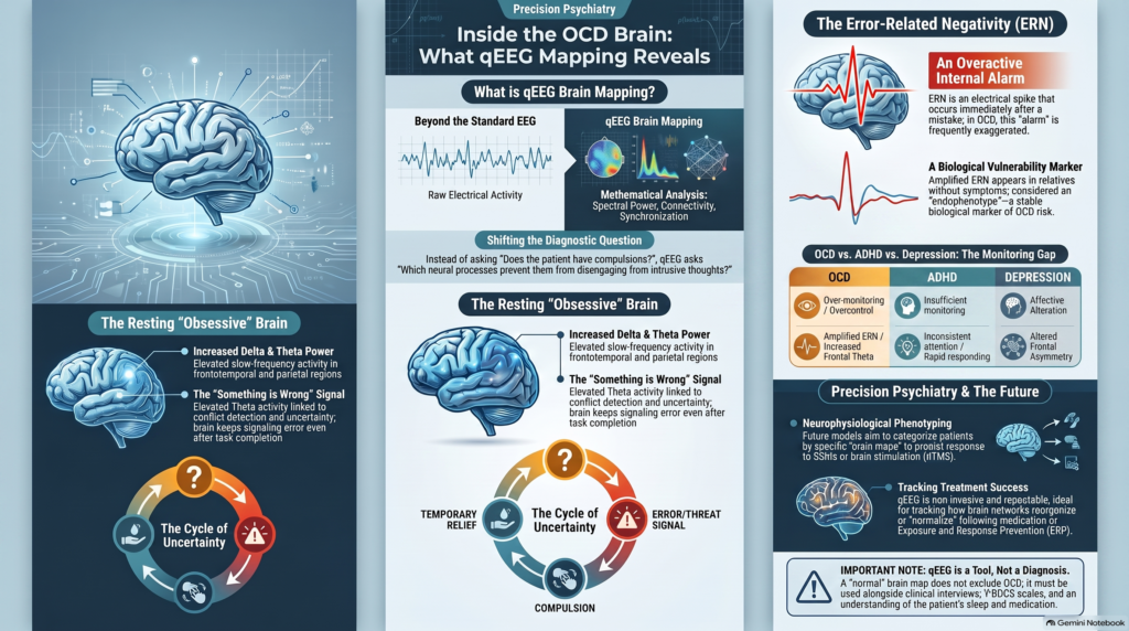
What Is qEEG Brain Mapping?
A conventional EEG records electrical activity generated by neuronal populations through electrodes placed on the scalp.
Quantitative EEG, or qEEG, mathematically analyses the digital EEG signal.
It can measure:
Spectral power
How much activity occurs within different frequency bands?
Relative power
What proportion of overall EEG activity is represented by delta, theta, alpha or beta activity?
Asymmetry
Are corresponding regions of the two hemispheres functioning differently?
Coherence and connectivity
How strongly are different brain regions synchronising their activity?
Source localisation
Where is particular electrical activity likely to originate?
Network organisation
How are different cortical regions interacting as functional systems?
The broader qEEG literature describes quantitative EEG as mathematical analysis of digital EEG capable of extracting frequency-band, complexity, connectivity and network information.
For OCD, these techniques allow us to move beyond the question:
“Does this patient have obsessions and compulsions?”
toward:
“What neural processes may be contributing to the inability to disengage from intrusive thoughts and repetitive behaviour?”
OCD Is Increasingly Viewed as a Brain-Network Disorder
For decades, neurobiological models of OCD have concentrated particularly on cortico-striato-thalamo-cortical circuits, often abbreviated as CSTC circuits.
These systems involve interactions among areas including:
- orbitofrontal cortex
- anterior cingulate cortex
- dorsolateral prefrontal cortex
- striatum
- thalamus
- supplementary motor regions
But newer research suggests that OCD cannot be reduced to a single loop.
Networks involved in:
- salience
- cognitive control
- error detection
- attention
- habit formation
- emotional processing
- behavioural inhibition
all appear relevant.
This is important for qEEG because EEG is especially good at measuring rapid changes in network activity and synchronisation.
What Does Resting qEEG Show in OCD?
One of the more informative recent resting-state EEG studies compared 25 people with OCD with 27 healthy controls.
The researchers found:
Increased delta power
particularly involving frontotemporal and parietal regions.
Increased theta power
particularly involving left frontotemporal regions.
Reduced functional connectivity in the delta range
when connectivity was measured using coherence.
Importantly, the study did not find significant group differences across every frequency band, illustrating that OCD cannot be reduced to one universally abnormal EEG pattern.
In simplified form, one electrophysiological pattern emerging from the literature is:
More slow-frequency activity + altered network communication
But the biological interpretation is more interesting than the simple map.
1. Increased Theta Activity in OCD
Theta activity broadly occupies approximately 4–8 Hz.
Theta oscillations are involved in several functions highly relevant to OCD:
- cognitive control
- conflict detection
- uncertainty processing
- error monitoring
- behavioural adjustment
- working memory
Several EEG studies have reported increased theta activity in OCD.
The 2023 resting-state study found elevated theta power particularly in the left frontotemporal region compared with healthy controls.
This fits an interesting neurobiological concept.
A person with OCD may not simply experience an intrusive thought.
The brain may continue signalling:
“Something is still wrong.”
even after the person has checked.
The “Something Is Not Right” Problem
Consider someone with contamination OCD.
They wash their hands.
Logically, they understand that their hands are clean.
But subjectively:
“It still doesn’t feel right.”
So they wash again.
Or consider checking OCD.
The person locks the door.
They remember locking it.
Yet an internal error signal persists:
“But what if I didn’t?”
They check again.
This cycle can be conceptualised as:
uncertainty
↓
error/threat signal
↓
compulsion
↓
temporary relief
↓
return of uncertainty
↓
repeat
One of the most fascinating findings in OCD electrophysiology is that abnormal performance and error monitoring can actually be measured.
2. Error-Related Negativity: One of the Strongest Electrophysiological Findings in OCD
Resting qEEG tells us what the brain is doing while relatively inactive.
But OCD may become even more interesting when the brain is required to perform a task and detect mistakes.
This introduces:
Error-Related Negativity — ERN
ERN is an event-related electrical potential that appears very rapidly after a person makes an error.
It is thought to arise largely from systems involving the medial frontal cortex and anterior cingulate cortex.
In healthy individuals, it functions somewhat like an internal error alarm:
“You made a mistake. Adjust your behaviour.”
In OCD, this error-monitoring response is frequently exaggerated.
A landmark study found increased error-related brain activity not only in people with OCD but also in their unaffected first-degree relatives.
That finding is particularly interesting because the relatives did not necessarily have OCD symptoms.
It raises the possibility that excessive error-monitoring activity may represent an endophenotype—a biological vulnerability characteristic associated with OCD rather than simply a consequence of having severe symptoms.
What Might an Excessive Error Signal Feel Like?
This offers an intriguing bridge between neurophysiology and clinical symptoms.
Most people lock their car and walk away.
The brain essentially says:
“Task completed.”
Someone with checking OCD may experience:
“Are you certain?”
They check again.
Then:
“But did you really check properly?”
Again.
The problem may therefore not simply be poor memory.
It may involve failure of the internal completion or certainty signal.
This does not mean ERN explains every compulsion.
But it provides one of the clearest examples of an apparently subjective psychiatric experience having a measurable electrophysiological correlate.
ERN Is Not the Same as Resting qEEG
This distinction is important.
Resting qEEG
examines spontaneous brain activity.
Event-related potentials
measure electrical responses linked to specific events or cognitive tasks.
Therefore a future comprehensive electrophysiological OCD assessment may extend beyond a static resting brain map.
It could involve:
Resting EEG
qEEG spectral analysis
connectivity
task EEG
error-monitoring ERPs
That would provide considerably more information about OCD neurophysiology than a single coloured qEEG map.
3. Delta Activity in OCD
The recent resting-state study also demonstrated increased delta oscillatory power in OCD, particularly involving frontotemporal and parietal regions.
Delta is generally a slower frequency range, approximately 1–4 Hz.
Excessive slow-wave activity during waking states can reflect altered cortical regulation, although its meaning is strongly dependent upon:
- brain region
- alertness
- medication
- sleep
- age
- recording conditions
Therefore:
Increased delta does not mean OCD.
But when increased slow activity repeatedly appears in particular networks across well-controlled OCD studies, it becomes biologically interesting.
The same recent study found both increased slow-frequency power and reduced delta coherence, suggesting that the abnormality may involve not only how strongly neurons oscillate but also how effectively regions coordinate their activity.
4. Functional Connectivity in OCD
The brain does not function as a collection of isolated electrodes.
A major advantage of modern qEEG is the ability to investigate:
Functional connectivity
This asks:
How strongly is activity in one region related to activity elsewhere?
One older EEG study specifically examined connectivity in OCD using source localisation and coherence analysis in drug-naïve patients, demonstrating abnormalities in the organisation of cortical networks.
Another resting-state study found reduced delta-band coherence across a network containing:
- frontocentral regions
- temporal regions
- parietal regions.
The precise connectivity findings across studies have not always been consistent.
That is important.
The safest conclusion is therefore:
OCD is associated with abnormal network organisation, but there is not yet one universally reproducible OCD connectivity map.
Why Connectivity May Ultimately Matter More Than Power
A traditional qEEG report might say:
“Theta is elevated.”
A network-based interpretation asks something much deeper:
“Which regions are failing to communicate normally while the brain processes uncertainty, detects errors or tries to inhibit repetitive behaviour?”
That question may eventually have greater clinical value.
OCD is unlikely to result from one brain region simply being “overactive.”
Instead, the disorder may involve abnormal communication between systems controlling error detection, attention, habits and behavioural inhibition.
5. Frontal Slowing and Frontal Asymmetry
Electrophysiological research has repeatedly highlighted frontal abnormalities in OCD.
Reported findings include:
- frontal slowing
- frontal asymmetry
- increased slow-wave activity
- altered frontal connectivity
These findings fit with the major role of frontal networks in:
- cognitive control
- inhibition
- decision-making
- uncertainty evaluation
- behavioural flexibility
But again, frontal slowing is not specific to OCD.
Similar changes may occur with:
- depression
- ADHD
- sleep deprivation
- medication effects
- neurological conditions
The value lies in identifying a pattern of abnormalities that makes sense within the clinical phenotype.
OCD Is Not Simply “Too Much Frontal Activity”
Popular neuroscience often oversimplifies OCD by saying that certain frontal regions are simply “overactive.”
The reality is more complicated.
A brain region can display:
- increased metabolic activity
- altered oscillatory power
- increased connectivity with one network
- reduced connectivity with another network
at the same time.
Therefore:
Increased activity does not necessarily mean improved function.
Nor does reduced activity necessarily mean impaired function.
What matters is appropriate regulation within the network.
6. Frontal-Midline Theta and Cognitive Control
Frontal-midline theta is especially interesting because it is closely associated with situations involving:
- conflict
- uncertainty
- error detection
- need for behavioural adjustment
These are precisely the cognitive states repeatedly encountered in OCD.
Imagine the obsessive question:
“Did I switch the gas off?”
The person checks.
But uncertainty remains.
The cognitive-control system continues evaluating:
Was the action correct?
Is further checking required?
Could something bad happen?
Persistent recruitment of error- and uncertainty-processing networks may contribute to the characteristic inability to reach a subjective sense of completion.
OCD May Be a Disorder of Over-Monitoring
One useful electrophysiological conceptualisation of OCD is:
The brain may monitor performance too strongly rather than too weakly.
This distinguishes OCD from conditions in which performance monitoring can be reduced.
The amplified ERN found in patients with OCD and unaffected relatives supports the idea that excessive internal error signalling may represent a relatively stable vulnerability marker rather than simply an effect of momentary anxiety.
This may help explain why reassurance often fails in OCD.
The patient may intellectually understand the reassurance.
Yet the internal error signal remains active.
Why Reassurance Often Does Not Work
Suppose someone repeatedly asks:
“Are you absolutely sure I won’t become infected?”
A family member answers:
“Yes.”
Anxiety temporarily falls.
But the brain generates another possibility:
“What if they misunderstood the question?”
Reassurance is sought again.
This is why repeatedly providing certainty can inadvertently function like a compulsion.
The goal of treatment is therefore not necessarily to make uncertainty disappear.
It is often to help the person learn:
“I can tolerate uncertainty without performing the compulsion.”
This is the principle behind Exposure and Response Prevention (ERP).
qEEG Does Not Replace ERP
This point is critical.
Understanding the neurobiology of OCD does not change the fact that evidence-based treatment remains essential.
NIMH identifies treatment approaches including psychotherapy, medication and brain-stimulation approaches, particularly in more resistant illness.
For psychotherapy, Exposure and Response Prevention is a central evidence-based treatment.
Brain mapping should therefore not become:
qEEG instead of ERP.
A more useful model is:
Better biological characterisation + evidence-based treatment.
Can qEEG Identify Different Types of OCD?
This may ultimately be one of the most useful applications.
OCD itself is heterogeneous.
Patients may predominantly experience:
- contamination and washing
- checking
- intrusive aggressive thoughts
- sexual obsessions
- religious obsessions
- symmetry and ordering
- “just-right” experiences
- responsibility fears
- mental compulsions
- reassurance seeking
There is no reason to assume that all of these phenotypes must have identical electrophysiology.
Indeed, early qEEG research suggested the possibility of distinct electrophysiological OCD subtypes.
An Important Early qEEG Treatment Study
A particularly interesting study examined 20 non-depressed patients with OCD treated with paroxetine.
Eighteen responded to treatment.
Among responders, qEEG subtype membership predicted SSRI response in 94.4% of cases. Patients in the responder subtype showed relatively strong baseline alpha activity, which reduced toward a more normal pattern following successful treatment.
This result is fascinating.
But it should be interpreted cautiously.
The study was:
- small
- conducted more than two decades ago
- based on a specific neurometric classification system
- not sufficient to establish qEEG-guided SSRI prescribing as routine clinical practice
Therefore we should not currently tell a patient:
“Your qEEG says paroxetine will work.”
But the study illustrates something important:
Physiological subtyping may eventually help predict treatment response.
This Is Where Precision Psychiatry Becomes Interesting
The traditional approach is:
Diagnosis: OCD
↓
Choose an evidence-based treatment
↓
Wait
↓
Measure response
But imagine a future model:
Diagnosis
clinical phenotype
symptom dimensions
qEEG phenotype
error-monitoring profile
connectivity pattern
↓
More individualised treatment selection
That is not yet routine practice.
But it represents a realistic research direction.
qEEG and Treatment-Resistant OCD
Treatment-resistant OCD is particularly relevant to neurophysiology.
When symptoms remain severe despite adequate trials of:
- serotonergic medication
- augmentation strategies
- structured ERP
additional biological approaches may become relevant.
NIMH specifically notes ongoing investigation of brain stimulation treatment for OCD, including research focused on treatment-resistant illness.
This creates an important potential role for EEG:
Can electrophysiology identify dysfunctional networks?
Can it provide baseline measurements before neuromodulation?
Can changes in those networks be tracked after treatment?
These questions are increasingly being investigated.
qEEG and Deep TMS for OCD
Deep transcranial magnetic stimulation (dTMS) represents an important development in treatment-resistant OCD.
A study examining patients undergoing dTMS recorded qEEG before and after treatment and found measurable electrophysiological changes alongside clinical improvement.
The importance of this research is not that qEEG has already become a routine method for selecting dTMS protocols.
Rather, it demonstrates that:
Treatment-induced changes in OCD may be measurable electrophysiologically.
This opens the possibility of eventually using qEEG to investigate:
- baseline network state
- biological treatment response
- target engagement
- longitudinal change
From Brain Mapping to Brain-Guided Neuromodulation
This may become one of the most interesting future applications of electrophysiology.
Traditional neuromodulation uses standardised stimulation targets.
Future approaches may increasingly ask:
Which network is abnormal in this patient?
Which oscillation is abnormal?
Which cortical region should be targeted?
Did stimulation actually modify the intended network?
This could combine:
qEEG
source localisation
connectivity analysis
TMS
clinical outcome measurement
That represents a more sophisticated form of precision interventional psychiatry.
OCD Versus Anxiety on qEEG
OCD is frequently mistaken for an anxiety disorder because anxiety is often prominent.
But the core pathology involves more than anxiety.
A person may experience:
intrusive thought
↓
error or threat signal
↓
urge to neutralise
↓
compulsion
The compulsion reduces anxiety temporarily, reinforcing the cycle.
Generalised anxiety may instead involve persistent worry without the same structured obsession-compulsion sequence.
qEEG cannot independently distinguish the two conditions, but the strong interest in error-monitoring electrophysiology in OCD provides an important additional dimension beyond general cortical arousal.
OCD Versus ADHD: Almost Opposite Error-Monitoring Problems?
This is another fascinating research question.
Both disorders can involve impaired executive functioning.
But their cognitive styles can appear very different.
ADHD
may involve:
- impulsive responding
- insufficient monitoring
- inconsistent attention
- rapid responding
OCD
may involve:
- excessive monitoring
- repeated checking
- overcontrol
- difficulty terminating evaluation
Electrophysiological research has found particularly robust increased error-related activity in OCD, supporting the concept of exaggerated performance monitoring.
This is an excellent example of why two psychiatric disorders that both affect executive function can have very different underlying neurophysiology.
OCD Versus Depression
Depression commonly accompanies OCD.
This matters enormously for qEEG interpretation.
Depression itself can alter:
- alpha activity
- theta activity
- frontal asymmetry
- coherence
Therefore, an OCD brain map cannot be interpreted without asking:
Is significant depression also present?
The recent resting-state OCD study specifically attempted to characterise slow-frequency and connectivity abnormalities, but the broader literature remains heterogeneous partly because psychiatric comorbidity can influence EEG results.
This is why diagnosis-first interpretation is essential.
Medication Can Alter EEG
Another critical issue is pharmacotherapy.
Patients with OCD may be taking:
- SSRIs
- clomipramine
- antipsychotic augmentation
- benzodiazepines
- sleep medication
- other psychotropic drugs
These medications can alter cortical electrical activity.
Therefore, a qEEG interpretation should ideally document:
- current medication
- dose
- duration
- caffeine
- sleep
- recent medication changes
Without this context, one risks interpreting a medication effect as an OCD biomarker.
Sleep Matters Too
OCD can cause substantial sleep disturbance.
Patients may:
- perform rituals late into the night
- remain mentally preoccupied
- repeatedly check before sleeping
- experience intrusive thoughts in bed
Sleep deprivation itself alters EEG activity.
It may increase slow-wave activity and impair attention.
Therefore, increased theta or delta activity must always be interpreted in relation to the patient’s sleep.
qEEG Brain Maps Are Not Pictures of Obsessions
Colour brain maps can appear remarkably convincing.
A patient may see red areas and assume:
“That is where my OCD is.”
That is not how qEEG should be interpreted.
Colours represent numerical measurements such as:
- spectral power
- relative power
- Z-scores
- coherence
- asymmetry
Red does not necessarily mean:
“diseased.”
Blue does not necessarily mean:
“underactive.”
The meaning depends entirely upon the parameter displayed.
Artifact Control Is Essential
qEEG is extremely sensitive to non-brain electrical activity.
Examples include:
- eye movements
- blinking
- jaw tension
- forehead muscle activity
- movement
- poor electrode contact
This becomes particularly important for higher-frequency activity.
The broader qEEG literature emphasises that biological state, equipment, electrodes and artifacts can substantially influence quantitative EEG interpretation.
Sophisticated software cannot rescue poor-quality EEG.
Can qEEG Diagnose OCD?
No—not currently.
OCD remains a clinical diagnosis based upon:
- obsessions
- compulsions
- distress
- time consumption
- impairment
- differential diagnosis
Research has demonstrated reproducible electrophysiological differences at a group level, particularly involving:
- slow-frequency activity
- frontal physiology
- functional connectivity
- error monitoring
But there is substantial overlap between individuals.
A normal brain map therefore does not exclude OCD.
And an abnormal map does not confirm it.
Can qEEG Measure OCD Severity?
Not reliably by itself.
Clinical severity is better measured using structured clinical assessment and instruments such as the Yale-Brown Obsessive Compulsive Scale (Y-BOCS).
qEEG can provide a physiological dimension.
Y-BOCS tells us:
How severe are the symptoms?
qEEG asks:
What electrophysiological abnormalities accompany them?
They answer different questions.
Can qEEG Tell Which Medicine Will Work?
Not yet reliably.
The early paroxetine study showing qEEG subtype-associated response is scientifically intriguing, but the dataset was far too small to establish a general prescribing rule.
At present, pharmacological treatment should continue to depend upon:
- diagnosis
- severity
- previous treatments
- medication tolerance
- comorbidity
- treatment response
- safety considerations
The future possibility is EEG-informed treatment prediction, not EEG-determined prescribing.
Can qEEG Track Treatment?
Potentially, and this may be more useful than diagnosis.
Because EEG is:
- non-invasive
- repeatable
- relatively inexpensive
- capable of millisecond-level measurement
the same patient can potentially be assessed longitudinally.
For example:
Baseline
Y-BOCS + qEEG
↓
Treatment
Medication + ERP
↓
Clinical reassessment
Y-BOCS
↓
Repeat electrophysiology
qEEG
Researchers can then ask:
Did symptom improvement occur alongside normalisation or reorganisation of specific brain networks?
This approach may be particularly interesting for:
- treatment-resistant OCD
- neurofeedback research
- TMS
- other neuromodulatory interventions
The Future: Resting qEEG Alone Will Probably Not Be Enough
The strongest future electrophysiological assessment of OCD may integrate several measurements.
Resting-state qEEG
Spectral power and connectivity.
Task-related EEG
What happens when uncertainty or conflict is introduced?
ERN
How strongly does the brain react to mistakes?
Frontal-midline theta
How does cognitive-control circuitry respond?
Connectivity analysis
How effectively are control networks interacting?
Clinical symptom dimensions
What kind of OCD does the patient actually have?
AI-based pattern recognition
Can combinations of features identify meaningful biological subtypes?
This is far more sophisticated than looking for one abnormal frequency.
From “OCD Brain Map” to Neurophysiological Phenotype
A better way to conceptualise qEEG is:
Patient A
Contamination OCD
- severe avoidance
- increased frontal slow activity
Patient B
Checking OCD
- extreme doubt
- exaggerated error-monitoring response
Patient C
OCD + ADHD
- mixed attention and control abnormalities
Patient D
OCD + depression
- additional affective electrophysiological abnormalities
Patient E
Treatment-resistant OCD
- network abnormality potentially relevant to neuromodulation research
These examples are conceptual.
They are not established clinical qEEG subtypes.
But they illustrate where the science could ultimately move:
From diagnostic labels toward neurophysiological phenotyping.
AI Could Change qEEG in OCD
A conventional EEG contains an enormous amount of data.
Potential features include:
- delta power
- theta power
- alpha power
- beta power
- regional asymmetry
- coherence
- phase relationships
- entropy
- signal complexity
- connectivity
- task responses
- ERN amplitude
A human cannot intuitively integrate hundreds of these variables.
Machine-learning models potentially can.
Rather than asking:
“Is theta increased?”
future models may analyse:
theta + delta + alpha + connectivity + ERN + clinical phenotype + treatment history
simultaneously.
This may eventually help classify:
- OCD physiological subtypes
- likely treatment responders
- treatment-resistant presentations
- neuromodulation candidates
That is one of the more plausible pathways toward precision psychiatry.
A 360-Degree Approach to OCD Assessment
qEEG should therefore sit within a broader clinical framework.
A comprehensive OCD assessment can include:
Detailed psychiatric interview
Identify obsessions, compulsions, avoidance and mental rituals.
OCD severity measurement
Quantify symptoms and impairment.
Assessment for common comorbidities
Including:
- depression
- ADHD
- tic disorders
- autism
- anxiety disorders
- substance use
- sleep disturbance
Evaluation of bipolarity where relevant
Particularly before complex pharmacological treatment.
qEEG / brain mapping
When there is a meaningful neurophysiological question.
ERP-focused psychological assessment
Identify the obsession → anxiety → compulsion → relief cycle.
Integrated treatment
Medication and psychotherapy should be designed around the patient’s actual clinical phenotype.
Brain Mapping and OCD Treatment in Chennai
My approach to OCD is based on diagnosis first, objective assessment where useful, and evidence-based treatment rather than technology for its own sake.
For selected patients, assessment can incorporate:
Detailed OCD diagnostic evaluation
Y-BOCS and structured symptom assessment
Evaluation for ADHD, depression, autism and other comorbidities
qEEG / brain mapping where clinically appropriate
Evidence-based pharmacotherapy
Exposure and Response Prevention
For patients requiring psychological intervention, structured OCD-focused therapy and ERP can be integrated with psychiatric management.
The goal is not merely:
“Reduce anxiety.”
It is to understand and treat the entire obsessive-compulsive cycle:
Intrusion → uncertainty → distress → compulsion → temporary relief → reinforcement
Frequently Asked Questions
Can a brain map diagnose OCD?
No. OCD remains a clinical diagnosis. qEEG can demonstrate electrophysiological abnormalities associated with OCD, but no qEEG pattern currently has sufficient sensitivity and specificity to establish the diagnosis independently.
What qEEG pattern is commonly reported in OCD?
Recent resting-state research has found increased delta and theta oscillatory power, particularly in frontotemporal and parietal regions, together with reduced delta-band coherence in one connectivity analysis.
What is the most interesting EEG biomarker in OCD?
One of the strongest candidates is increased error-related negativity (ERN)—an exaggerated electrophysiological response immediately after making an error. Increased error-related activity has also been demonstrated in unaffected first-degree relatives, suggesting it may represent a vulnerability marker.
Does increased theta prove OCD?
No. Increased theta activity occurs in multiple psychiatric, neurological and physiological states. Its distribution, connectivity, recording quality and clinical context must all be considered.
Can qEEG choose the correct OCD medicine?
Not currently. Early studies suggested that qEEG subtypes might predict SSRI response, but these results require much larger prospective replication before being used routinely for medication selection.
Can qEEG be used before and after treatment?
Yes, EEG is non-invasive and repeatable. Longitudinal research has examined electrophysiological changes after pharmacological treatment and brain-stimulation interventions, but routine clinical interpretation of those changes is still developing.
Is qEEG the same as an ERP?
No.
qEEG generally quantifies ongoing EEG activity.
Event-related potentials measure electrical responses associated with particular events or cognitive tasks.
ERN is an ERP associated with making errors.
Is brain mapping painful?
No. EEG electrodes record naturally occurring electrical activity from the scalp. They do not send electricity into the brain.
OCD Brain Mapping and Precision Psychiatry
Dr. Srinivas Rajkumar T
MBBS (Madurai Medical College), MD (AIIMS New Delhi), DNB, MBA (BITS Pilani)
Senior Consultant Psychiatrist
Apollo Clinic, Velachery, Chennai
Opposite Phoenix Marketcity
Brain mapping should not be marketed as a coloured scan that simply says “OCD present.”
Its scientific potential is considerably more interesting.
The future of OCD assessment may therefore move beyond simply identifying obsessions and compulsions toward understanding the neurophysiological phenotype underlying them.
That is where qEEG may eventually make its greatest contribution.
Selected References
- Perera MPN, Mallawaarachchi S, Bailey NW, Murphy OW, Fitzgerald PB. Obsessive-compulsive disorder is associated with increased electroencephalographic delta and theta oscillatory power but reduced delta connectivity. 2023.
- Velikova S, Locatelli M, Insacco C, Smeraldi E, Comi G, Leocani L. Dysfunctional brain circuitry in obsessive-compulsive disorder: source and coherence analysis of EEG rhythms. NeuroImage. 2010;49:977–983. doi:10.1016/j.neuroimage.2009.08.015.
- Riesel A, Endrass T, Kaufmann C, Kathmann N. Overactive error-related brain activity as a candidate endophenotype for obsessive-compulsive disorder. Am J Psychiatry. 2011.
- Hansen ES, Prichep LS, Bolwig TG, John ER. Quantitative electroencephalography in OCD patients treated with paroxetine. Clin Electroencephalogr. 2003;34(2):70–74.
- Popa LL, Dragos H, Pantelemon C, Rosu OV, Strilciuc S. The Role of Quantitative EEG in the Diagnosis of Neuropsychiatric Disorders. J Med Life. 2020;13(1):8–15. doi:10.25122/jml-2019-0085.