Brain Mapping in Autism Spectrum Disorder: qEEG, Connectivity and the Neurophysiology of the Autistic Brain
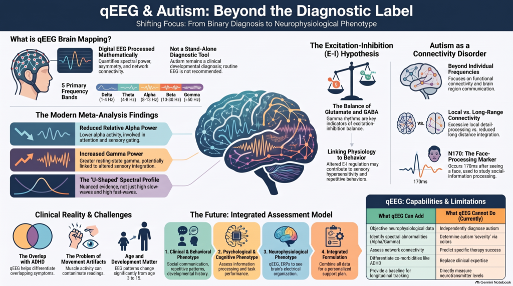 Can autism be seen on a brain map?
Autism Spectrum Disorder (ASD) is a neurodevelopmental condition, meaning that differences in brain development and information processing are central to the condition. This naturally raises an important question:
If autism involves differences in brain function, can we measure some of those differences objectively?
One increasingly studied approach is quantitative electroencephalography (qEEG), commonly called brain mapping.
qEEG measures and mathematically analyses the electrical activity generated by the brain. Research in autism has identified differences involving:
- alpha activity
- theta activity
- beta and gamma activity
- functional connectivity
- hemispheric and regional organisation
- sensory-processing responses
- social-information processing
- mu rhythms
- large-scale neural-network dynamics
But an important distinction must be made at the outset:
There is currently no single qEEG pattern that diagnoses autism.
Autism remains a clinical developmental diagnosis. Current paediatric guidance does not recommend routine EEG simply to establish an autism diagnosis; conventional EEG is generally reserved for situations in which epilepsy or another specific neurological indication is suspected.
The more scientifically appropriate question is therefore not:
“Does the brain map prove autism?”
but:
“What does this individual’s electrophysiological profile tell us about their brain function?”
That distinction is central to the emerging concept of objective neurodevelopmental assessment.
What Is qEEG Brain Mapping?
A conventional EEG records tiny fluctuations in electrical potential generated predominantly by synchronised cortical neuronal activity.
qEEG takes this digital EEG recording and applies mathematical analysis to quantify characteristics such as:
Spectral power
How much activity exists within different frequency ranges?
Relative power
What proportion of total EEG activity lies within each frequency band?
Peak frequencies
At what frequencies does an individual brain generate its dominant rhythms?
Asymmetry
Do corresponding regions of the two hemispheres behave differently?
Coherence and connectivity
How synchronised are different brain regions?
Network organisation
How efficiently are different cortical systems interacting?
The qEEG review supplied for this series describes quantitative EEG as digital EEG processed mathematically to examine frequency bands, signal complexity, connectivity and network characteristics. It specifically notes that qEEG has been widely investigated in autism and that quantitative markers may be associated with differences in brain function.
The same review importantly concludes that qEEG should generally provide additional objective information alongside other investigations rather than function as an immediate stand-alone diagnosis.
Why Is EEG Particularly Interesting in Autism?
Autism is extraordinarily heterogeneous.
Two autistic individuals can differ substantially in:
- language
- intelligence
- attention
- sensory processing
- social communication
- repetitive behaviour
- motor coordination
- adaptive functioning
- emotional regulation
- sleep
- associated ADHD symptoms
- epilepsy risk
This heterogeneity creates a problem for biomarker research.
If autism represents multiple developmental pathways that eventually produce overlapping behavioural characteristics, there may be no single universal “autism brain wave.”
That is increasingly what the EEG literature suggests.
Instead of one diagnostic signature, researchers are finding multiple electrophysiological dimensions and subgroups. Large meta-analytic studies also report significant methodological and individual variability between autism EEG studies.
This makes qEEG potentially more interesting for phenotyping autism than simply attempting to diagnose it.
The Major EEG Frequency Bands
Brain electrical activity can be broadly divided into frequency ranges.
Delta
Approximately 1–4 Hz
Theta
Approximately 4–8 Hz
Alpha
Approximately 8–13 Hz
Beta
Approximately 13–30 Hz
Gamma
Above approximately 30 Hz
These bands should not be interpreted as individual “functions.” Each reflects complex neuronal dynamics, and the significance of a frequency depends on:
- brain region
- developmental age
- behavioural state
- eyes-open versus eyes-closed recording
- task being performed
- medication
- sleep
- technical recording parameters
Nevertheless, differences in their distribution can provide useful information about brain-network physiology.
The Historical “U-Shaped” qEEG Pattern in Autism
Earlier autism EEG literature proposed an intriguing pattern:
Increased activity at the slower end of the spectrum
Delta / theta ↑
combined with:
Reduced middle-frequency activity
Alpha ↓
and:
Increased faster-frequency activity
Beta / gamma ↑
This became known as a possible “U-shaped” spectral profile of autism.
Earlier reviews reported evidence supporting such a pattern, while also emphasising substantial inconsistency between studies.
However, newer evidence has refined this picture considerably.
What Does the Modern Meta-Analysis Show?
A large systematic review and meta-analysis published in Translational Psychiatry synthesised resting-state EEG power studies in autistic and neurotypical participants across the conventional frequency bands.
Two of the clearer group-level findings were:
Reduced relative alpha power
and
Increased gamma power
in autistic participants compared with neurotypical comparison groups.
The alpha difference produced a moderate effect, while gamma differences were also prominent.
This is important because it demonstrates both the promise and the limitation of EEG biomarkers.
There are reproducible physiological differences at a population level.
But there is substantial overlap between individuals.
Therefore:
An individual with reduced alpha does not automatically have autism.
And:
An autistic individual does not necessarily demonstrate the “classic” qEEG pattern.
1. Alpha Activity in Autism
Of all resting-state EEG findings, alpha activity has become particularly interesting.
Alpha oscillations are prominent during quiet wakefulness and are strongly involved in:
- regulation of cortical excitability
- attention
- sensory gating
- internal versus external information processing
- coordination between brain networks
The modern meta-analysis found reduced relative resting-state alpha power in autism, although considerable variation exists between studies and individuals.
This could potentially reflect differences in how autistic brains regulate cortical processing.
But alpha should not simply be interpreted as:
“Low alpha = autism.”
A more useful assessment examines:
- absolute alpha power
- relative alpha power
- regional distribution
- alpha peak frequency
- frontal versus posterior alpha
- alpha connectivity
Individual Alpha Peak Frequency
Another potentially valuable measurement is the individual alpha peak frequency.
Instead of assuming that every person’s alpha rhythm behaves identically between 8 and 13 Hz, qEEG can identify where an individual’s dominant alpha rhythm actually peaks.
This matters particularly in neurodevelopment.
Brain oscillatory patterns change considerably with:
- age
- maturation
- intellectual functioning
- attention
- arousal
Therefore, interpreting a child’s EEG using adult assumptions can be misleading.
This is one reason age-adjusted interpretation is essential in paediatric qEEG.
2. Gamma Activity: One of the Most Interesting Autism Findings
Gamma oscillations represent faster-frequency brain activity and are involved in processes such as:
- sensory integration
- perceptual binding
- attention
- local cortical processing
- communication within neuronal networks
The 2023 meta-analysis found greater absolute and relative resting-state gamma power in autistic individuals compared with neurotypical controls.
This finding is particularly interesting because gamma rhythms depend heavily on interactions between excitatory and inhibitory neuronal circuits.
That has generated considerable interest in the possibility that some forms of autism involve altered excitation–inhibition regulation.
The Excitation–Inhibition Hypothesis
Normal brain function requires a carefully regulated balance between neuronal excitation and inhibition.
Broadly:
Glutamatergic systems
contribute substantially to excitation.
GABAergic systems
contribute substantially to inhibition.
One influential hypothesis proposes that altered regulation of these systems may contribute to features of autism such as:
- sensory hypersensitivity
- sensory filtering difficulties
- repetitive behaviour
- atypical cortical synchronisation
- altered perceptual processing
Gamma oscillations are particularly relevant because inhibitory interneuron networks play an important role in their generation.
However, scalp qEEG does not directly measure glutamate or GABA.
Therefore it would be inappropriate to look at increased gamma on a brain map and conclude:
“This patient has a GABA deficiency.”
EEG can generate hypotheses about network physiology. It does not directly measure neurotransmitter concentrations.
3. Theta Activity in Autism
Theta oscillations are involved in processes including:
- attention
- memory
- cognitive control
- learning
- long-range coordination between brain regions
Some autism studies have reported increased theta activity, while others have found reduced or unchanged theta power.
For example, work comparing ASD, ADHD, combined ASD+ADHD and neurotypical groups found that children with autism-related diagnoses showed reduced theta and alpha power relative to groups without autism, illustrating how EEG findings depend strongly on the population being studied.
This is an excellent example of why autism cannot be reduced to a single frequency abnormality.
Autism Versus ADHD on qEEG
This becomes especially relevant clinically because ADHD frequently coexists with autism.
Both may present with:
- distractibility
- impulsivity
- executive difficulties
- sensory-seeking behaviour
- restlessness
- difficulties completing tasks
But the underlying developmental phenotype may differ considerably.
ADHD research historically concentrated heavily on theta/beta relationships.
Autism EEG research has increasingly focused on:
- alpha activity
- gamma activity
- sensory processing
- connectivity
- social-information processing
- developmental trajectories
Studies directly comparing autistic children, children with ADHD and children with both conditions demonstrate that overlapping clinical symptoms do not necessarily imply identical electrophysiology.
This is where multidimensional assessment becomes particularly interesting.
A Child Can Have Autism AND ADHD
Another reason simplistic brain-map diagnosis fails is that neurodevelopmental conditions frequently coexist.
A patient may have:
Autism
ADHD
Anxiety
Sleep disturbance
and potentially:
Learning or intellectual difficulties.
Each may influence EEG activity.
Therefore, the qEEG should be interpreted within a clinical formulation rather than asking whether the map is simply:
“Autistic” or “not autistic.”
4. Autism as a Connectivity Disorder
Perhaps the most important direction in modern autism electrophysiology is functional connectivity.
The brain operates through networks.
Normal social communication requires efficient coordination between systems involved in:
- visual perception
- facial recognition
- language
- attention
- emotional processing
- motor imitation
- memory
- reward
- executive control
qEEG can estimate relationships between signals recorded from different cortical regions.
These measures include:
- coherence
- phase synchronisation
- amplitude coupling
- functional connectivity
- graph-theoretical network measures
Local Overconnectivity and Long-Range Underconnectivity?
One influential theory proposed that autism involves:
excessive local connectivity
combined with:
reduced long-distance connectivity.
This was intuitively attractive because it could potentially explain why some autistic individuals demonstrate:
- exceptional detail-focused processing
while simultaneously experiencing difficulty integrating information across complex social environments.
However, modern evidence suggests that this model is too simple.
A systematic evaluation of EEG and MEG connectivity studies found substantial variability according to:
- developmental stage
- brain region
- frequency band
- analysis technique
- recording paradigm
The literature does support atypical connectivity in autism, but not a universal rule that every autistic brain has local overconnectivity and long-range underconnectivity.
Connectivity May Be More Important Than Individual Frequencies
Traditional qEEG might say:
“Alpha is reduced.”
Network analysis asks:
“Which regions are communicating differently?”
That may ultimately be much more clinically informative.
For example, two autistic individuals could have similar total alpha power while having very different patterns of:
- frontal–temporal connectivity
- interhemispheric synchronisation
- posterior network organisation
- sensory network connectivity
Recent resting-state research continues to demonstrate altered theta and alpha connectivity in young autistic children, although sample sizes remain small and findings require replication.
This is likely to become an increasingly important area of qEEG research.
5. Sensory Processing and qEEG
Sensory differences are highly relevant in autism.
Autistic individuals may experience unusual sensitivity or reduced sensitivity to:
- sound
- light
- touch
- clothing textures
- movement
- smell
- taste
- crowded environments
These experiences are not simply behavioural phenomena.
Sensory processing depends on how neural networks:
- detect incoming information,
- filter irrelevant information,
- determine salience,
- integrate information across sensory systems,
- regulate behavioural responses.
EEG is particularly well suited to studying these processes because neural responses occur within milliseconds.
Resting qEEG provides one layer of information.
Task-related EEG and event-related potentials (ERPs) provide another.
Beyond qEEG: ERPs and Autism
When a particular stimulus is repeatedly presented—for example a face, sound or visual pattern—the electrical brain response can be averaged.
This produces an event-related potential.
Several ERP components have attracted autism research interest.
Among the most important are:
- P1
- N170
- visual evoked responses
- auditory evoked responses
The Autism Biomarkers Consortium for Clinical Trials has evaluated several EEG measures as potential autism biomarkers. Candidate measures including the P1 and N170 responses to faces and visual-evoked-potential measures demonstrated moderate short-term stability, an important requirement if biomarkers are eventually to be used in clinical trials or longitudinal assessment.
This illustrates an important evolution:
From resting brain maps alone
toward:
Resting EEG + task EEG + ERP + connectivity.
The N170 and Face Processing
The N170 is an EEG response occurring approximately 170 milliseconds after presentation of a visual stimulus and is particularly associated with face processing.
Because social perception and interpretation of facial information are important areas of autism research, the N170 has become a prominent candidate biomarker.
However, the goal is not to create a simple:
“N170 autism test.”
Rather, measures such as N170 may help researchers quantify aspects of social-information processing objectively.
This could eventually become useful for:
- biological stratification
- longitudinal monitoring
- clinical trials
- measuring response to targeted interventions
The Autism Biomarkers Consortium work represents an important movement toward systematically validating such electrophysiological measures rather than relying on isolated small studies.
6. Mu Rhythm and Autism
Another fascinating EEG signal is the mu rhythm.
Mu activity generally occurs around the alpha-frequency range over sensorimotor cortex.
It typically changes when a person:
- performs an action
- observes another person performing an action
- engages certain motor and social-perception networks
This generated considerable interest in autism because imitation, action observation and social learning involve sensorimotor networks.
Researchers therefore investigated whether abnormalities of mu suppression might provide information about autism-related social processing.
The results have been interesting but heterogeneous.
Mu rhythm is therefore better viewed as a research window into sensorimotor and social-network physiology than as a diagnostic autism marker.
qEEG and Neurofeedback in Autism
Mu rhythm also became one target for EEG neurofeedback.
Neurofeedback works by measuring selected aspects of brain activity and providing real-time feedback that allows the individual to practise changing that activity.
Small studies have examined mu-based neurofeedback in autistic children, including research demonstrating that participants could learn to modify mu activity alongside changes in selected behavioural outcomes.
More recently, a 2024 randomised, placebo-controlled study involving 60 autistic children aged 3–6 years compared active mu-rhythm neurofeedback with sham neurofeedback alongside behavioural interventions. The study adds to evidence that EEG-based closed-loop interventions are technically feasible and potentially useful, but one trial does not establish neurofeedback as a standard treatment for autism.
The appropriate interpretation at present is:
Promising adjunctive technology—still under active investigation.
Not:
Established replacement for developmental and behavioural intervention.
Brain Mapping Should Guide Questions, Not Create Labels
Perhaps the biggest conceptual mistake in qEEG is looking at a colour map and saying:
“This is an autistic brain.”
The more scientifically sophisticated questions are:
Is the alpha rhythm developmentally appropriate?
Is there unusual slow or fast-frequency activity?
Are network connectivity patterns atypical?
Is there evidence of a separate attentional phenotype?
Are there electrophysiological features relevant to sensory processing?
Is epilepsy or epileptiform activity a concern?
Is there a measurable baseline that might be useful longitudinally?
Those are much more useful questions.
Why Colour Maps Can Be Misleading
qEEG maps frequently display activity using colours such as:
red – yellow – green – blue
But red does not mean:
“Abnormal autism area.”
A colour generally represents a numerical value—for example:
- power
- relative power
- statistical deviation
- connectivity
- asymmetry
Interpretation depends on:
- the parameter being displayed
- reference montage
- normative database
- age
- electrode quality
- eye state
- alertness
- artifact removal
- medications
- developmental level
The uploaded qEEG review specifically emphasises the importance of biological variability, equipment, electrodes, waking state and artifacts in interpreting quantitative EEG.
Movement Artifact Is Particularly Important in Autism Assessments
Recording clean EEG in children with autism can be challenging.
Potential sources of artifact include:
- movement
- blinking
- jaw clenching
- facial-muscle activity
- touching electrodes
- intolerance of the cap or sensors
- difficulty remaining still
These factors can especially contaminate faster-frequency activity such as beta and gamma.
Therefore, a visually dramatic gamma abnormality should never be interpreted without first asking:
Is this neuronal activity—or muscle artifact?
Recording quality is as important as the sophistication of the software analysing it.
qEEG and Epilepsy in Autism: A Different Clinical Question
Autism is also associated with an increased prevalence of epilepsy and epileptiform EEG abnormalities.
This creates an important distinction between:
Routine clinical EEG
used when seizures or epileptic syndromes are suspected
and
qEEG brain mapping
used to quantify aspects of brain electrical activity.
Current clinical guidance does not recommend obtaining EEG routinely merely because someone is autistic. EEG is appropriate when the clinical history raises concern for epilepsy or a relevant neurological syndrome.
This is particularly important if there is:
- suspected seizure activity
- unexplained episodes of altered awareness
- unusual nocturnal events
- developmental regression
- clinically concerning episodic behaviour
In such situations, conventional neurological EEG assessment may take priority over qEEG brain mapping.
Can qEEG Diagnose Autism?
Not currently.
Research demonstrates group-level differences between autistic and neurotypical populations, but no qEEG marker currently possesses sufficient sensitivity, specificity and reproducibility to replace clinical developmental diagnosis.
The strongest recent resting-state meta-analysis demonstrated meaningful group differences—particularly reduced relative alpha and increased gamma—but also substantial heterogeneity across studies.
This is exactly what would be expected from a condition called a spectrum.
Can a Normal qEEG Exclude Autism?
No.
An autistic person can have a resting EEG that falls largely within population reference ranges.
Conversely, an abnormal qEEG does not establish autism because similar abnormalities can appear in:
- ADHD
- epilepsy
- intellectual disability
- anxiety
- sleep deprivation
- medication effects
- other neurodevelopmental conditions
qEEG therefore adds information—it does not replace developmental assessment.
Perhaps We Are Asking the Wrong Diagnostic Question
Instead of asking:
“Can EEG diagnose autism?”
the future question may be:
“Can EEG identify biologically meaningful subtypes within autism?”
That is potentially much more useful.
Imagine two children who both fulfil behavioural criteria for autism.
Child A
Prominent sensory hypersensitivity
- relatively intact language
- significant gamma abnormalities
Child B
Marked language delay
- attentional impairment
- altered alpha organisation
Child C
Autism + ADHD
- a different arousal profile
Child D
Autism + epilepsy
- additional epileptiform abnormalities
These examples are conceptual rather than validated qEEG subtypes.
But they illustrate why neurophysiological phenotyping may eventually matter more than attempting to produce a binary autism EEG test.
Autism Is Developmental: Age Matters Enormously
A child’s brain is continuously changing.
EEG characteristics evolve with:
- infancy
- childhood
- adolescence
- adulthood
Therefore, an EEG abnormality cannot be interpreted without considering developmental age.
This may partly explain why autism EEG studies sometimes appear contradictory.
A pattern detectable at age 3 may not look identical at age 15.
Recent longitudinal work examining early childhood EEG trajectories has again found developmental differences in resting-state power between autistic and typically developing children, reinforcing the importance of analysing trajectories rather than single static measurements.
This is an important direction for future research.
Longitudinal Brain Mapping May Be More Valuable Than a Single Test
Rather than asking whether one EEG is “positive for autism,” repeated measurements could potentially examine:
baseline physiology
↓
developmental intervention
↓
repeat neurophysiological assessment
↓
change in spectral power or connectivity
↓
correlation with functional improvement.
EEG is particularly attractive for this type of research because it is:
- non-invasive
- repeatable
- relatively portable
- capable of high temporal resolution
The challenge is ensuring sufficiently standardised recording conditions for meaningful comparisons.
qEEG and Artificial Intelligence
This may ultimately transform the field.
A conventional qEEG generates many variables.
For multiple electrodes, algorithms can calculate:
- delta power
- theta power
- alpha power
- beta power
- gamma power
- spectral ratios
- asymmetry
- connectivity
- phase relationships
- entropy
- complexity
- network topology
The number of possible features rapidly becomes too large for simple visual inspection.
Machine learning can potentially identify multidimensional patterns that humans cannot easily recognise manually.
Research is increasingly combining EEG—and in some projects fNIRS—with machine-learning approaches to investigate autism classification and physiological phenotyping. The field remains developmental, however, and performance in research datasets should not automatically be interpreted as equivalent to clinical diagnostic accuracy in real-world populations.
The Future May Be EEG + fNIRS + Cognitive Assessment
EEG measures:
Electrical neuronal dynamics.
Functional near-infrared spectroscopy—fNIRS—measures:
Changes in cortical oxygenation and haemodynamics.
These modalities provide complementary information.
Future neurodevelopmental assessment may therefore combine:
Developmental history
Behavioural assessment
Cognitive testing
EEG/qEEG
ERP
fNIRS
machine learning
rather than searching endlessly for one test capable of diagnosing autism.
Emerging literature is already examining combined EEG/fNIRS approaches together with machine learning for autism research.
qEEG Should Not Replace Good Autism Assessment
The strongest autism assessment remains multidimensional.
Depending on age and presentation, evaluation should address:
Social communication
How does the individual initiate and sustain reciprocal interaction?
Restricted and repetitive patterns
Are repetitive behaviour, focused interests or insistence on sameness present?
Sensory characteristics
Are there unusual sensory sensitivities or sensory-seeking behaviours?
Developmental history
Were the characteristics present during the developmental period?
Language and cognition
Are there speech, language, intellectual or learning difficulties?
Adaptive functioning
How independently does the individual function in everyday life?
Comorbidity
Is ADHD, anxiety, sleep disturbance, intellectual disability or another condition present?
Only after understanding the phenotype does a physiological investigation become clinically meaningful.
A More Objective Autism Assessment Model
A comprehensive neurodevelopmental assessment can therefore be conceptualised as:
Clinical developmental phenotype
What characteristics are present?
↓
Psychological and cognitive phenotype
How does the person process information and perform?
↓
Behavioural phenotype
How do these differences affect daily functioning?
↓
Neurophysiological phenotype
What does EEG/qEEG demonstrate?
↓
Integrated formulation
What combination of supports and interventions is appropriate?
This avoids both extremes:
relying entirely on subjective impressions
and
claiming that technology can replace clinical expertise.
What qEEG Cannot Currently Tell Us
qEEG cannot independently determine:
- whether someone definitely has autism
- whether a child will later develop autism
- the “severity” of autism from map colours
- intellectual ability
- language prognosis
- which therapy will definitely work
- whether an individual requires medication
- whether neurofeedback will work
- the exact neurotransmitter abnormality underlying symptoms
And importantly:
There is no scientifically validated single “autism brain map.”
The qEEG literature instead points toward substantial electrophysiological heterogeneity.
What qEEG Can Potentially Add
Used appropriately, brain mapping can contribute several useful dimensions.
Objective neurophysiological information
EEG records measurable brain electrical activity rather than relying entirely on behavioural observation.
Identification of electrophysiological differences
Spectral abnormalities involving alpha, gamma and other frequencies may help characterise the individual physiological phenotype.
Connectivity assessment
qEEG allows researchers to examine how different regions communicate.
Separation of overlapping neurodevelopmental questions
Autism, ADHD, intellectual disability, sleep problems and epilepsy may coexist. Physiological assessment may add another dimension to clinical differentiation.
Baseline measurement
A baseline EEG may be useful when longitudinal physiological assessment or research monitoring is planned.
Neurofeedback research
EEG can potentially identify and train selected oscillatory patterns, although clinical evidence in autism remains developing.
Brain Mapping in Autism: Where the Science Is Heading
The first generation of qEEG research asked:
“Which EEG frequency is abnormal in autism?”
The second generation asked:
“Which brain regions are connected differently?”
The next generation is beginning to ask:
“Can we identify reproducible neurophysiological subtypes that predict cognition, sensory function, developmental trajectory or treatment response?”
That is a much more powerful question.
The future of objective autism assessment is unlikely to consist of one red-and-blue brain map.
It is more likely to involve:
**EEG spectral power
- connectivity
- developmental trajectories
- ERPs
- cognitive testing
- behavioural phenotype
- AI-based pattern recognition**
Brain Mapping and Autism Assessment in Chennai
For children, adolescents or adults undergoing assessment for possible autism, my approach is to use clinical diagnosis first and technology as an additional source of objective information where appropriate.
A comprehensive evaluation may incorporate:
Detailed developmental and psychiatric assessment
Autism-focused clinical evaluation
Assessment for ADHD and other neurodevelopmental conditions
Cognitive and psychological assessment where indicated
qEEG / brain mapping when it can answer a meaningful clinical question
Integrated treatment and support planning
The objective is not simply to produce a colourful map.
It is to understand the individual’s developmental, cognitive, behavioural and neurophysiological profile as comprehensively as possible.
Frequently Asked Questions
Can brain mapping diagnose autism?
No. Autism remains a clinical developmental diagnosis. qEEG can provide objective electrophysiological information but is not currently sufficiently accurate or specific to diagnose autism independently. Current clinical guidance also does not recommend routine EEG solely to establish an autism diagnosis.
What is the most consistent qEEG finding in autism?
There is no universal pattern. However, a recent systematic review and meta-analysis found reduced relative resting-state alpha power and increased gamma power among the more reproducible group-level findings.
Does autism cause increased theta?
Some studies have reported increased slow-wave activity, but the literature is inconsistent. Direct comparisons of autism, ADHD and combined presentations have demonstrated different theta patterns, highlighting the heterogeneity of autism electrophysiology.
Is there an “autism EEG signature”?
Not yet. There are group-level differences in spectral power and connectivity, but substantial overlap exists between autistic and neurotypical individuals.
What does low alpha mean in autism?
Reduced relative alpha power has been demonstrated at the group level. Alpha is involved in regulation of cortical processing and network coordination, but low alpha alone does not diagnose autism.
What does high gamma mean?
Increased gamma power has been reported in autism and may be relevant to differences in local cortical processing and excitation–inhibition regulation. However, scalp gamma is also especially vulnerable to muscle artifact and cannot directly measure neurotransmitter abnormalities.
Can qEEG identify autism versus ADHD?
Not reliably by itself. The two conditions can coexist and demonstrate overlapping electrophysiology. A combined developmental, psychiatric and objective cognitive assessment is more informative.
Is EEG required in every autistic child?
No. Conventional EEG is generally indicated when there is a clinical reason to suspect epilepsy or another neurological disorder rather than being performed routinely simply because autism is present.
Can neurofeedback treat autism?
EEG neurofeedback is an interesting emerging adjunct. Mu-rhythm and other protocols have produced encouraging findings in small studies, and placebo-controlled research continues to develop. It should currently be regarded as an adjunctive and investigational approach rather than a replacement for established developmental, educational, speech-language or behavioural interventions.
Autism, qEEG and the Future of Precision Neurodevelopmental Psychiatry
Dr. Srinivas Rajkumar T
MBBS (Madurai Medical College), MD (AIIMS New Delhi), DNB, MBA (BITS Pilani)
Senior Consultant Psychiatrist
Apollo Clinic, Velachery, Chennai
Opposite Phoenix Marketcity
My clinical approach focuses on diagnosis-first, evidence-based psychiatry with objective assessment wherever it adds meaningful information.
qEEG should not be presented as a machine that “detects autism.”
Its more important future role may be much deeper:
Clinical assessment tells us the developmental phenotype.
Cognitive testing tells us how the individual processes information.
qEEG tells us about the electrical organisation of the brain.
Connectivity analysis tells us how different brain regions interact.
Longitudinal measurement may eventually tell us how these systems change with development and intervention.
The real promise of brain mapping in autism is therefore not replacing clinical diagnosis.
It is helping psychiatry move from a broad label of “Autism Spectrum Disorder” toward a much more individual understanding of the person’s neurodevelopmental phenotype.
Selected References
- Popa LL, Dragos H, Pantelemon C, Rosu OV, Strilciuc S. The Role of Quantitative EEG in the Diagnosis of Neuropsychiatric Disorders. J Med Life. 2020;13(1):8–15. doi:10.25122/jml-2019-0085.
- Billeci L, Sicca F, Maharatna K, et al. On the application of quantitative EEG for characterizing autistic brain: a systematic review. Front Hum Neurosci. 2013;7:442.
- Neo WS, Foti D, Keehn B, Kelleher BL. Resting-state EEG power differences in autism spectrum disorder: a systematic review and meta-analysis. Transl Psychiatry. 2023;13:389.
- O’Reilly C, Lewis JD, Elsabbagh M. Is functional brain connectivity atypical in autism? A systematic review of EEG and MEG studies. PLoS One. 2017.
- Webb SJ, et al. The Autism Biomarkers Consortium for Clinical Trials: Initial evaluation of a battery of candidate EEG biomarkers. Am J Psychiatry. 2023.
- Friedrich EVC, Sivanathan A, Lim T, et al. An effective neurofeedback intervention to improve social interactions in children with Autism Spectrum Disorder. J Autism Dev Disord. 2015;45:4084–4100. doi:10.1007/s10803-015-2523-5.
- Wang XN, Zhang T, Han BC, et al. Wearable EEG neurofeedback based-on machine learning algorithms for children with autism: a randomized, placebo-controlled study. Curr Med Sci. 2024;44:1141–1147. doi:10.1007/s11596-024-2938-3.